{"title":"通过全身静脉取样和68Ga-DOTATOC正电子发射断层扫描定位肿瘤诱导的骨软化症。","authors":"Tomomi Nakao, Ken Takeshima, Shuhei Morita, Ichiro Yamauchi, Sho Koyasu, Taka-Aki Matsuoka","doi":"10.1210/jcemcr/luaf012","DOIUrl":null,"url":null,"abstract":"<p><p>Tumor-induced osteomalacia is characterized by hypophosphatemia and fragility fractures caused by fibroblast growth factor 23 (FGF23)-producing tumors. We report a case of tumor-induced osteomalacia in which the tumor location could be determined by gallium 68 (<sup>68</sup>Ga)-DOTATOC positron emission tomography (PET)/computed tomography (CT). A 74-year-old woman had recurrent fractures and bone pain. Blood tests showed hypophosphatemia and elevated serum alkaline phosphatase and FGF23 levels and CT and bone scintigraphy showed multiple bone fractures. Tumor-induced osteomalacia was therefore suspected. Indium 111 (<sup>111</sup>In)-pentetreotide scintigraphy showed focus of increased activity in the head, and CT and magnetic resonance images showed a mass-like lesion in the posterior ethmoidal sinus. However, in systemic venous sampling, serum FGF23 level was highest in the left common iliac vein. <sup>68</sup>Ga-DOTATOC PET/CT clearly demonstrated focal uptake in the left anterior inferior iliac spine consistent with systemic venous sampling. Retrospectively analyzed, focal uptake in the head was considered to be a physiological uptake in the pituitary gland. The tumor was resected and the pathological diagnosis was phosphaturic mesenchymal tumor. A combination of systemic venous sampling and <sup>68</sup>Ga-DOTATOC PET/CT was useful in detection of a small FGF23-producing tumor. Precise tumor localization in such cases requires careful interpretation of scintigraphy.</p>","PeriodicalId":73540,"journal":{"name":"JCEM case reports","volume":"3 2","pages":"luaf012"},"PeriodicalIF":0.0000,"publicationDate":"2025-01-24","publicationTypes":"Journal Article","fieldsOfStudy":null,"isOpenAccess":false,"openAccessPdf":"https://www.ncbi.nlm.nih.gov/pmc/articles/PMC11758137/pdf/","citationCount":"0","resultStr":"{\"title\":\"Tumor-Induced Osteomalacia Localized by Systemic Venous Sampling and <sup>68</sup>Ga-DOTATOC Positron Emission Tomography.\",\"authors\":\"Tomomi Nakao, Ken Takeshima, Shuhei Morita, Ichiro Yamauchi, Sho Koyasu, Taka-Aki Matsuoka\",\"doi\":\"10.1210/jcemcr/luaf012\",\"DOIUrl\":null,\"url\":null,\"abstract\":\"<p><p>Tumor-induced osteomalacia is characterized by hypophosphatemia and fragility fractures caused by fibroblast growth factor 23 (FGF23)-producing tumors. We report a case of tumor-induced osteomalacia in which the tumor location could be determined by gallium 68 (<sup>68</sup>Ga)-DOTATOC positron emission tomography (PET)/computed tomography (CT). A 74-year-old woman had recurrent fractures and bone pain. Blood tests showed hypophosphatemia and elevated serum alkaline phosphatase and FGF23 levels and CT and bone scintigraphy showed multiple bone fractures. Tumor-induced osteomalacia was therefore suspected. Indium 111 (<sup>111</sup>In)-pentetreotide scintigraphy showed focus of increased activity in the head, and CT and magnetic resonance images showed a mass-like lesion in the posterior ethmoidal sinus. However, in systemic venous sampling, serum FGF23 level was highest in the left common iliac vein. <sup>68</sup>Ga-DOTATOC PET/CT clearly demonstrated focal uptake in the left anterior inferior iliac spine consistent with systemic venous sampling. Retrospectively analyzed, focal uptake in the head was considered to be a physiological uptake in the pituitary gland. The tumor was resected and the pathological diagnosis was phosphaturic mesenchymal tumor. A combination of systemic venous sampling and <sup>68</sup>Ga-DOTATOC PET/CT was useful in detection of a small FGF23-producing tumor. Precise tumor localization in such cases requires careful interpretation of scintigraphy.</p>\",\"PeriodicalId\":73540,\"journal\":{\"name\":\"JCEM case reports\",\"volume\":\"3 2\",\"pages\":\"luaf012\"},\"PeriodicalIF\":0.0000,\"publicationDate\":\"2025-01-24\",\"publicationTypes\":\"Journal Article\",\"fieldsOfStudy\":null,\"isOpenAccess\":false,\"openAccessPdf\":\"https://www.ncbi.nlm.nih.gov/pmc/articles/PMC11758137/pdf/\",\"citationCount\":\"0\",\"resultStr\":null,\"platform\":\"Semanticscholar\",\"paperid\":null,\"PeriodicalName\":\"JCEM case reports\",\"FirstCategoryId\":\"1085\",\"ListUrlMain\":\"https://doi.org/10.1210/jcemcr/luaf012\",\"RegionNum\":0,\"RegionCategory\":null,\"ArticlePicture\":[],\"TitleCN\":null,\"AbstractTextCN\":null,\"PMCID\":null,\"EPubDate\":\"2025/2/1 0:00:00\",\"PubModel\":\"eCollection\",\"JCR\":\"\",\"JCRName\":\"\",\"Score\":null,\"Total\":0}","platform":"Semanticscholar","paperid":null,"PeriodicalName":"JCEM case reports","FirstCategoryId":"1085","ListUrlMain":"https://doi.org/10.1210/jcemcr/luaf012","RegionNum":0,"RegionCategory":null,"ArticlePicture":[],"TitleCN":null,"AbstractTextCN":null,"PMCID":null,"EPubDate":"2025/2/1 0:00:00","PubModel":"eCollection","JCR":"","JCRName":"","Score":null,"Total":0}
Tumor-Induced Osteomalacia Localized by Systemic Venous Sampling and 68Ga-DOTATOC Positron Emission Tomography.
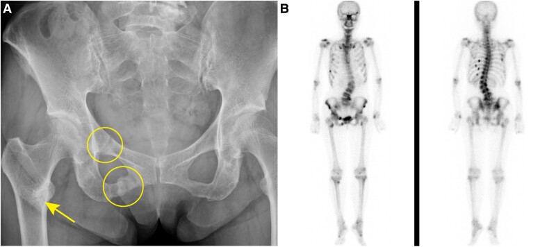
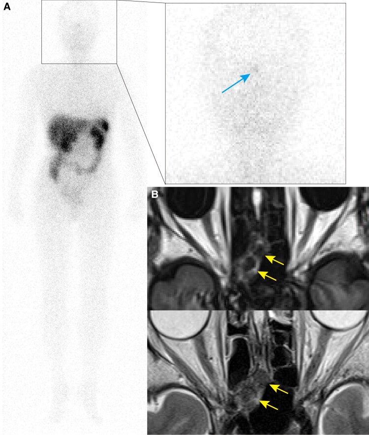
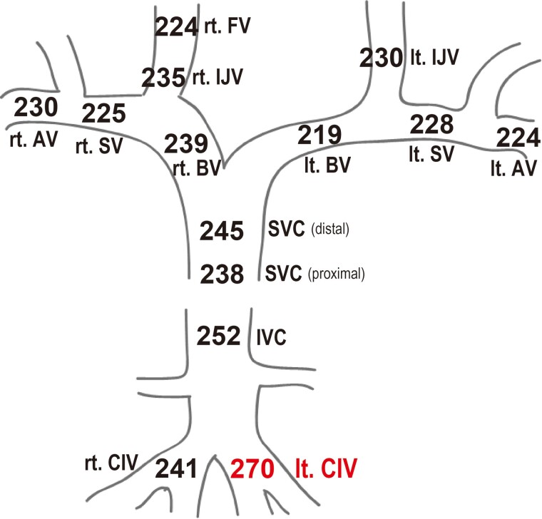
Tumor-induced osteomalacia is characterized by hypophosphatemia and fragility fractures caused by fibroblast growth factor 23 (FGF23)-producing tumors. We report a case of tumor-induced osteomalacia in which the tumor location could be determined by gallium 68 (68Ga)-DOTATOC positron emission tomography (PET)/computed tomography (CT). A 74-year-old woman had recurrent fractures and bone pain. Blood tests showed hypophosphatemia and elevated serum alkaline phosphatase and FGF23 levels and CT and bone scintigraphy showed multiple bone fractures. Tumor-induced osteomalacia was therefore suspected. Indium 111 (111In)-pentetreotide scintigraphy showed focus of increased activity in the head, and CT and magnetic resonance images showed a mass-like lesion in the posterior ethmoidal sinus. However, in systemic venous sampling, serum FGF23 level was highest in the left common iliac vein. 68Ga-DOTATOC PET/CT clearly demonstrated focal uptake in the left anterior inferior iliac spine consistent with systemic venous sampling. Retrospectively analyzed, focal uptake in the head was considered to be a physiological uptake in the pituitary gland. The tumor was resected and the pathological diagnosis was phosphaturic mesenchymal tumor. A combination of systemic venous sampling and 68Ga-DOTATOC PET/CT was useful in detection of a small FGF23-producing tumor. Precise tumor localization in such cases requires careful interpretation of scintigraphy.

 求助内容:
求助内容: 应助结果提醒方式:


