Chan Hong Moon, Frank S Lieberman, Hoby P Hetherington, Jullie W Pan
{"title":"人脑快速哈达玛编码7T光谱成像。","authors":"Chan Hong Moon, Frank S Lieberman, Hoby P Hetherington, Jullie W Pan","doi":"10.3390/tomography11010007","DOIUrl":null,"url":null,"abstract":"<p><p><b>Background/Objectives</b>: The increased SNR available at 7T combined with fast readout trajectories enables accelerated spectroscopic imaging acquisitions for clinical applications. In this report, we evaluate the performance of a Hadamard slice encoding strategy with a 2D rosette trajectory for multi-slice fast spectroscopic imaging at 7T. <b>Methods</b>: Moderate-TE (~40 ms) spin echo and J-refocused polarization transfer sequences were acquired with simultaneous Hadamard multi-slice excitations and rosette in-plane encoding. The moderate spin echo sequence, which targets singlet compounds (i.e., N-acetyl aspartate, creatine, and choline), uses cascaded multi-slice RF excitation pulses to minimize the chemical shift dispersion error. The J-refocused sequence targets coupled spin systems (i.e., glutamate and myo-inositol) using simultaneous multi-slice excitation to maintain the same TE across all slices. A modified Hadamard slice encoding strategy was used to decrease the peak RF pulse amplitude of the simultaneous multi-slice excitation pulse for the J-refocused acquisition. <b>Results</b>: The accuracy of multi-slice and single-slice rosette spectroscopic imaging (RSI) is comparable to conventional Cartesian-encoded spectroscopic imaging (CSI). Spectral analyses for the J-refocused studies of glutamate and myo-inositol show that the Cramer Rao lower bounds are not significantly different between the fast RSI and conventional CSI studies. Linear regressions of creatine/N-acetyl aspartate and glutamate/N-acetyl aspartate with tissue gray matter content are consistent with literature values. <b>Conclusions</b>: With minimal gradient demands and fast acquisition times, the 2.2 min to 9 min for single- to four-slice RSI acquisitions are well tolerated by healthy subjects and tumor patients, and show results that are consistent with clinical outcomes.</p>","PeriodicalId":51330,"journal":{"name":"Tomography","volume":"11 1","pages":""},"PeriodicalIF":2.2000,"publicationDate":"2025-01-13","publicationTypes":"Journal Article","fieldsOfStudy":null,"isOpenAccess":false,"openAccessPdf":"https://www.ncbi.nlm.nih.gov/pmc/articles/PMC11769540/pdf/","citationCount":"0","resultStr":"{\"title\":\"Fast Hadamard-Encoded 7T Spectroscopic Imaging of Human Brain.\",\"authors\":\"Chan Hong Moon, Frank S Lieberman, Hoby P Hetherington, Jullie W Pan\",\"doi\":\"10.3390/tomography11010007\",\"DOIUrl\":null,\"url\":null,\"abstract\":\"<p><p><b>Background/Objectives</b>: The increased SNR available at 7T combined with fast readout trajectories enables accelerated spectroscopic imaging acquisitions for clinical applications. In this report, we evaluate the performance of a Hadamard slice encoding strategy with a 2D rosette trajectory for multi-slice fast spectroscopic imaging at 7T. <b>Methods</b>: Moderate-TE (~40 ms) spin echo and J-refocused polarization transfer sequences were acquired with simultaneous Hadamard multi-slice excitations and rosette in-plane encoding. The moderate spin echo sequence, which targets singlet compounds (i.e., N-acetyl aspartate, creatine, and choline), uses cascaded multi-slice RF excitation pulses to minimize the chemical shift dispersion error. The J-refocused sequence targets coupled spin systems (i.e., glutamate and myo-inositol) using simultaneous multi-slice excitation to maintain the same TE across all slices. A modified Hadamard slice encoding strategy was used to decrease the peak RF pulse amplitude of the simultaneous multi-slice excitation pulse for the J-refocused acquisition. <b>Results</b>: The accuracy of multi-slice and single-slice rosette spectroscopic imaging (RSI) is comparable to conventional Cartesian-encoded spectroscopic imaging (CSI). Spectral analyses for the J-refocused studies of glutamate and myo-inositol show that the Cramer Rao lower bounds are not significantly different between the fast RSI and conventional CSI studies. Linear regressions of creatine/N-acetyl aspartate and glutamate/N-acetyl aspartate with tissue gray matter content are consistent with literature values. <b>Conclusions</b>: With minimal gradient demands and fast acquisition times, the 2.2 min to 9 min for single- to four-slice RSI acquisitions are well tolerated by healthy subjects and tumor patients, and show results that are consistent with clinical outcomes.</p>\",\"PeriodicalId\":51330,\"journal\":{\"name\":\"Tomography\",\"volume\":\"11 1\",\"pages\":\"\"},\"PeriodicalIF\":2.2000,\"publicationDate\":\"2025-01-13\",\"publicationTypes\":\"Journal Article\",\"fieldsOfStudy\":null,\"isOpenAccess\":false,\"openAccessPdf\":\"https://www.ncbi.nlm.nih.gov/pmc/articles/PMC11769540/pdf/\",\"citationCount\":\"0\",\"resultStr\":null,\"platform\":\"Semanticscholar\",\"paperid\":null,\"PeriodicalName\":\"Tomography\",\"FirstCategoryId\":\"3\",\"ListUrlMain\":\"https://doi.org/10.3390/tomography11010007\",\"RegionNum\":4,\"RegionCategory\":\"医学\",\"ArticlePicture\":[],\"TitleCN\":null,\"AbstractTextCN\":null,\"PMCID\":null,\"EPubDate\":\"\",\"PubModel\":\"\",\"JCR\":\"Q2\",\"JCRName\":\"RADIOLOGY, NUCLEAR MEDICINE & MEDICAL IMAGING\",\"Score\":null,\"Total\":0}","platform":"Semanticscholar","paperid":null,"PeriodicalName":"Tomography","FirstCategoryId":"3","ListUrlMain":"https://doi.org/10.3390/tomography11010007","RegionNum":4,"RegionCategory":"医学","ArticlePicture":[],"TitleCN":null,"AbstractTextCN":null,"PMCID":null,"EPubDate":"","PubModel":"","JCR":"Q2","JCRName":"RADIOLOGY, NUCLEAR MEDICINE & MEDICAL IMAGING","Score":null,"Total":0}
引用次数: 0
摘要
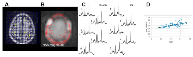
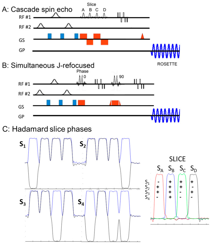
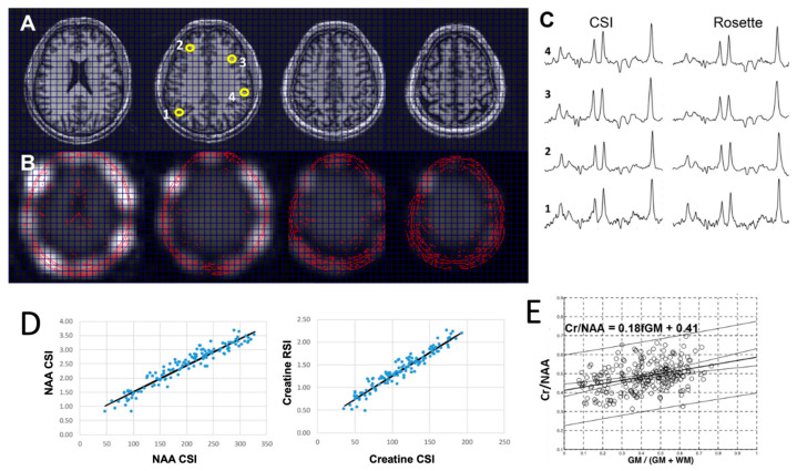
背景/目的:在7T时提高信噪比,结合快速读出轨迹,可以加速临床应用的光谱成像采集。在这篇报告中,我们评估了一种具有二维玫瑰花形轨迹的Hadamard切片编码策略在7T下的多片快速光谱成像的性能。方法:采用Hadamard多片同步激励和面内玫瑰花编码,获得中等te (~40 ms)自旋回波和j -再聚焦极化传递序列。针对单线态化合物(即n -乙酰天冬氨酸、肌酸和胆碱)的适度自旋回波序列,使用级联的多层射频激发脉冲来最小化化学位移色散误差。j -重聚焦序列靶向耦合自旋系统(即谷氨酸和肌醇),使用同时的多片激励来保持所有片上相同的TE。采用改进的Hadamard切片编码策略,降低了同时多片激励脉冲的射频脉冲峰值幅度,实现了j重聚焦采集。结果:多层和单层玫瑰花环光谱成像(RSI)的精度与传统的笛卡尔编码光谱成像(CSI)相当。j -refocus研究的谷氨酸和肌醇的光谱分析表明,快速RSI和常规CSI研究之间的Cramer Rao下限没有显著差异。肌酸/ n -乙酰天冬氨酸和谷氨酸/ n -乙酰天冬氨酸与组织灰质含量的线性回归与文献值一致。结论:健康受试者和肿瘤患者对单排至四排RSI采集的2.2 min至9 min具有较好的耐受性,且梯度要求最小,采集时间短,其结果与临床结果一致。
Fast Hadamard-Encoded 7T Spectroscopic Imaging of Human Brain.
Background/Objectives: The increased SNR available at 7T combined with fast readout trajectories enables accelerated spectroscopic imaging acquisitions for clinical applications. In this report, we evaluate the performance of a Hadamard slice encoding strategy with a 2D rosette trajectory for multi-slice fast spectroscopic imaging at 7T. Methods: Moderate-TE (~40 ms) spin echo and J-refocused polarization transfer sequences were acquired with simultaneous Hadamard multi-slice excitations and rosette in-plane encoding. The moderate spin echo sequence, which targets singlet compounds (i.e., N-acetyl aspartate, creatine, and choline), uses cascaded multi-slice RF excitation pulses to minimize the chemical shift dispersion error. The J-refocused sequence targets coupled spin systems (i.e., glutamate and myo-inositol) using simultaneous multi-slice excitation to maintain the same TE across all slices. A modified Hadamard slice encoding strategy was used to decrease the peak RF pulse amplitude of the simultaneous multi-slice excitation pulse for the J-refocused acquisition. Results: The accuracy of multi-slice and single-slice rosette spectroscopic imaging (RSI) is comparable to conventional Cartesian-encoded spectroscopic imaging (CSI). Spectral analyses for the J-refocused studies of glutamate and myo-inositol show that the Cramer Rao lower bounds are not significantly different between the fast RSI and conventional CSI studies. Linear regressions of creatine/N-acetyl aspartate and glutamate/N-acetyl aspartate with tissue gray matter content are consistent with literature values. Conclusions: With minimal gradient demands and fast acquisition times, the 2.2 min to 9 min for single- to four-slice RSI acquisitions are well tolerated by healthy subjects and tumor patients, and show results that are consistent with clinical outcomes.
TomographyMedicine-Radiology, Nuclear Medicine and Imaging
CiteScore
2.70
自引率
10.50%
发文量
222
期刊介绍:
TomographyTM publishes basic (technical and pre-clinical) and clinical scientific articles which involve the advancement of imaging technologies. Tomography encompasses studies that use single or multiple imaging modalities including for example CT, US, PET, SPECT, MR and hyperpolarization technologies, as well as optical modalities (i.e. bioluminescence, photoacoustic, endomicroscopy, fiber optic imaging and optical computed tomography) in basic sciences, engineering, preclinical and clinical medicine.
Tomography also welcomes studies involving exploration and refinement of contrast mechanisms and image-derived metrics within and across modalities toward the development of novel imaging probes for image-based feedback and intervention. The use of imaging in biology and medicine provides unparalleled opportunities to noninvasively interrogate tissues to obtain real-time dynamic and quantitative information required for diagnosis and response to interventions and to follow evolving pathological conditions. As multi-modal studies and the complexities of imaging technologies themselves are ever increasing to provide advanced information to scientists and clinicians.
Tomography provides a unique publication venue allowing investigators the opportunity to more precisely communicate integrated findings related to the diverse and heterogeneous features associated with underlying anatomical, physiological, functional, metabolic and molecular genetic activities of normal and diseased tissue. Thus Tomography publishes peer-reviewed articles which involve the broad use of imaging of any tissue and disease type including both preclinical and clinical investigations. In addition, hardware/software along with chemical and molecular probe advances are welcome as they are deemed to significantly contribute towards the long-term goal of improving the overall impact of imaging on scientific and clinical discovery.

 求助内容:
求助内容: 应助结果提醒方式:


