Philip Kyeremeh Jnr Oppong, Hiroyuki Hamaguchi, Maho Kitagawa, Nina Patzke, Kevin C Wakeman, Khin Khin Tha
{"title":"脊髓区髓磷脂MRI评价指标的比较敏感性。","authors":"Philip Kyeremeh Jnr Oppong, Hiroyuki Hamaguchi, Maho Kitagawa, Nina Patzke, Kevin C Wakeman, Khin Khin Tha","doi":"10.3390/tomography11010008","DOIUrl":null,"url":null,"abstract":"<p><p><b>Background/Objectives:</b> Although multiple magnetic resonance imaging (MRI) indices are known to be sensitive to the noninvasive assessment of myelin integrity, their relative sensitivities have not been directly compared. This study aimed to identify the most sensitive MRI index for characterizing myelin composition in the spinal cord's gray matter (GM) and white matter (WM). <b>Methods:</b> MRI was performed on a deer's ex vivo cervical spinal cord. Quantitative indices known to be sensitive to myelin, including the myelin water fraction (MWF), magnetization transfer ratio (MTR), the signal ratio between T1- and T2-weighted images (T1W/T2W), fractional anisotropy (FA), mean diffusivity (MD), electrical conductivity (σ), and T1, T2, and T1ρ relaxation times were calculated. Their mean values were compared using repeated measures analysis of variance (ANOVA) and post hoc Bonferroni tests or Friedman and post hoc Wilcoxon tests to identify differences across GM and WM columns possessing distinct myelin distributions, as revealed by histological analysis. Relationships among the indices were examined using Spearman's rank-order correlation analysis. Corrected <i>p</i> < 0.05 was considered statistically significant. <b>Results:</b> All indices except σ differed significantly between GM and all WM columns. Two of the three WM columns had significantly different MWF, FA, MD, and T2, whereas one WM column had significantly different MTR, σ, T1, and T1ρ from the others. A significant moderate to very strong correlation was observed among most indices. <b>Conclusions:</b> The sensitivity of MRI indices in distinguishing spinal cord regions varied. A strategic combination of two or more indices may allow the accurate differentiation of spinal cord regions.</p>","PeriodicalId":51330,"journal":{"name":"Tomography","volume":"11 1","pages":""},"PeriodicalIF":2.2000,"publicationDate":"2025-01-14","publicationTypes":"Journal Article","fieldsOfStudy":null,"isOpenAccess":false,"openAccessPdf":"https://www.ncbi.nlm.nih.gov/pmc/articles/PMC11769071/pdf/","citationCount":"0","resultStr":"{\"title\":\"Comparative Sensitivity of MRI Indices for Myelin Assessment in Spinal Cord Regions.\",\"authors\":\"Philip Kyeremeh Jnr Oppong, Hiroyuki Hamaguchi, Maho Kitagawa, Nina Patzke, Kevin C Wakeman, Khin Khin Tha\",\"doi\":\"10.3390/tomography11010008\",\"DOIUrl\":null,\"url\":null,\"abstract\":\"<p><p><b>Background/Objectives:</b> Although multiple magnetic resonance imaging (MRI) indices are known to be sensitive to the noninvasive assessment of myelin integrity, their relative sensitivities have not been directly compared. This study aimed to identify the most sensitive MRI index for characterizing myelin composition in the spinal cord's gray matter (GM) and white matter (WM). <b>Methods:</b> MRI was performed on a deer's ex vivo cervical spinal cord. Quantitative indices known to be sensitive to myelin, including the myelin water fraction (MWF), magnetization transfer ratio (MTR), the signal ratio between T1- and T2-weighted images (T1W/T2W), fractional anisotropy (FA), mean diffusivity (MD), electrical conductivity (σ), and T1, T2, and T1ρ relaxation times were calculated. Their mean values were compared using repeated measures analysis of variance (ANOVA) and post hoc Bonferroni tests or Friedman and post hoc Wilcoxon tests to identify differences across GM and WM columns possessing distinct myelin distributions, as revealed by histological analysis. Relationships among the indices were examined using Spearman's rank-order correlation analysis. Corrected <i>p</i> < 0.05 was considered statistically significant. <b>Results:</b> All indices except σ differed significantly between GM and all WM columns. Two of the three WM columns had significantly different MWF, FA, MD, and T2, whereas one WM column had significantly different MTR, σ, T1, and T1ρ from the others. A significant moderate to very strong correlation was observed among most indices. <b>Conclusions:</b> The sensitivity of MRI indices in distinguishing spinal cord regions varied. A strategic combination of two or more indices may allow the accurate differentiation of spinal cord regions.</p>\",\"PeriodicalId\":51330,\"journal\":{\"name\":\"Tomography\",\"volume\":\"11 1\",\"pages\":\"\"},\"PeriodicalIF\":2.2000,\"publicationDate\":\"2025-01-14\",\"publicationTypes\":\"Journal Article\",\"fieldsOfStudy\":null,\"isOpenAccess\":false,\"openAccessPdf\":\"https://www.ncbi.nlm.nih.gov/pmc/articles/PMC11769071/pdf/\",\"citationCount\":\"0\",\"resultStr\":null,\"platform\":\"Semanticscholar\",\"paperid\":null,\"PeriodicalName\":\"Tomography\",\"FirstCategoryId\":\"3\",\"ListUrlMain\":\"https://doi.org/10.3390/tomography11010008\",\"RegionNum\":4,\"RegionCategory\":\"医学\",\"ArticlePicture\":[],\"TitleCN\":null,\"AbstractTextCN\":null,\"PMCID\":null,\"EPubDate\":\"\",\"PubModel\":\"\",\"JCR\":\"Q2\",\"JCRName\":\"RADIOLOGY, NUCLEAR MEDICINE & MEDICAL IMAGING\",\"Score\":null,\"Total\":0}","platform":"Semanticscholar","paperid":null,"PeriodicalName":"Tomography","FirstCategoryId":"3","ListUrlMain":"https://doi.org/10.3390/tomography11010008","RegionNum":4,"RegionCategory":"医学","ArticlePicture":[],"TitleCN":null,"AbstractTextCN":null,"PMCID":null,"EPubDate":"","PubModel":"","JCR":"Q2","JCRName":"RADIOLOGY, NUCLEAR MEDICINE & MEDICAL IMAGING","Score":null,"Total":0}
Comparative Sensitivity of MRI Indices for Myelin Assessment in Spinal Cord Regions.
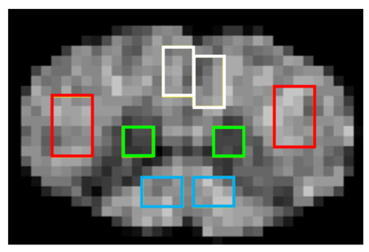
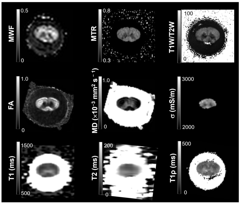
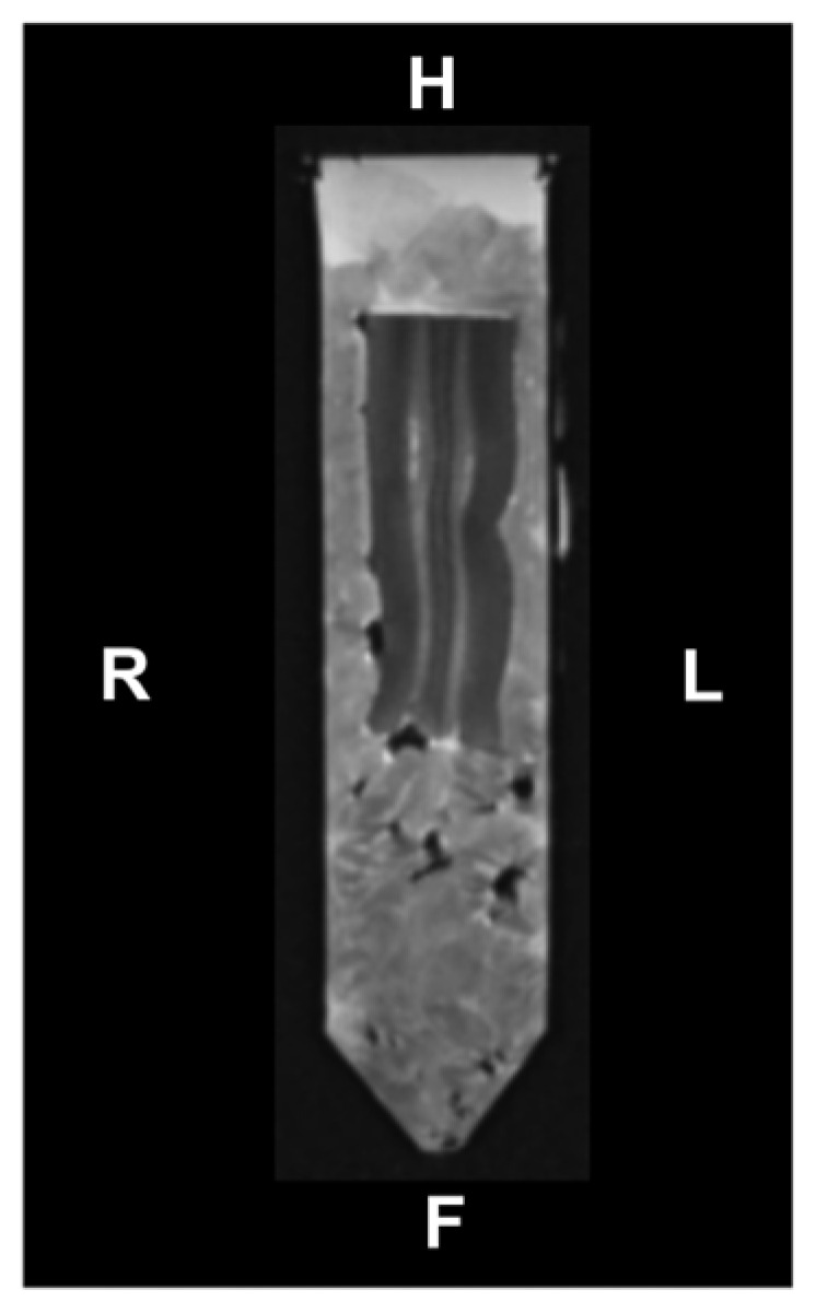
Background/Objectives: Although multiple magnetic resonance imaging (MRI) indices are known to be sensitive to the noninvasive assessment of myelin integrity, their relative sensitivities have not been directly compared. This study aimed to identify the most sensitive MRI index for characterizing myelin composition in the spinal cord's gray matter (GM) and white matter (WM). Methods: MRI was performed on a deer's ex vivo cervical spinal cord. Quantitative indices known to be sensitive to myelin, including the myelin water fraction (MWF), magnetization transfer ratio (MTR), the signal ratio between T1- and T2-weighted images (T1W/T2W), fractional anisotropy (FA), mean diffusivity (MD), electrical conductivity (σ), and T1, T2, and T1ρ relaxation times were calculated. Their mean values were compared using repeated measures analysis of variance (ANOVA) and post hoc Bonferroni tests or Friedman and post hoc Wilcoxon tests to identify differences across GM and WM columns possessing distinct myelin distributions, as revealed by histological analysis. Relationships among the indices were examined using Spearman's rank-order correlation analysis. Corrected p < 0.05 was considered statistically significant. Results: All indices except σ differed significantly between GM and all WM columns. Two of the three WM columns had significantly different MWF, FA, MD, and T2, whereas one WM column had significantly different MTR, σ, T1, and T1ρ from the others. A significant moderate to very strong correlation was observed among most indices. Conclusions: The sensitivity of MRI indices in distinguishing spinal cord regions varied. A strategic combination of two or more indices may allow the accurate differentiation of spinal cord regions.
TomographyMedicine-Radiology, Nuclear Medicine and Imaging
CiteScore
2.70
自引率
10.50%
发文量
222
期刊介绍:
TomographyTM publishes basic (technical and pre-clinical) and clinical scientific articles which involve the advancement of imaging technologies. Tomography encompasses studies that use single or multiple imaging modalities including for example CT, US, PET, SPECT, MR and hyperpolarization technologies, as well as optical modalities (i.e. bioluminescence, photoacoustic, endomicroscopy, fiber optic imaging and optical computed tomography) in basic sciences, engineering, preclinical and clinical medicine.
Tomography also welcomes studies involving exploration and refinement of contrast mechanisms and image-derived metrics within and across modalities toward the development of novel imaging probes for image-based feedback and intervention. The use of imaging in biology and medicine provides unparalleled opportunities to noninvasively interrogate tissues to obtain real-time dynamic and quantitative information required for diagnosis and response to interventions and to follow evolving pathological conditions. As multi-modal studies and the complexities of imaging technologies themselves are ever increasing to provide advanced information to scientists and clinicians.
Tomography provides a unique publication venue allowing investigators the opportunity to more precisely communicate integrated findings related to the diverse and heterogeneous features associated with underlying anatomical, physiological, functional, metabolic and molecular genetic activities of normal and diseased tissue. Thus Tomography publishes peer-reviewed articles which involve the broad use of imaging of any tissue and disease type including both preclinical and clinical investigations. In addition, hardware/software along with chemical and molecular probe advances are welcome as they are deemed to significantly contribute towards the long-term goal of improving the overall impact of imaging on scientific and clinical discovery.

 求助内容:
求助内容: 应助结果提醒方式:


