Yu Dong Ning, Yi Xuan Song, Yu Qin He, Han Li, Shao Yan Liu
{"title":"新辅助免疫治疗联合化疗后头颈部鳞状细胞癌影像学检查与手术病理的差异。","authors":"Yu Dong Ning, Yi Xuan Song, Yu Qin He, Han Li, Shao Yan Liu","doi":"10.14740/wjon1973","DOIUrl":null,"url":null,"abstract":"<p><strong>Background: </strong>We here investigated the value of imaging examination in evaluating tumor remission-based surgery in patients with head and neck squamous cell carcinoma (HNSCC), who had undergone neoadjuvant immunotherapy combined with chemotherapy (NICC).</p><p><strong>Methods: </strong>HNSCC patients who underwent NICC and surgery from May 2021 to September 2023 were retrospectively analyzed. All patients had to undergo imaging examination evaluation, including enhanced computed tomography (CT) and enhanced magnetic resonance (MR) imaging before and after NICC. Data related to clinical parameters, complete response of the primary site (PrCR), complete response of the primary site and the lymph node (PLCR), complete response of the lymph node (LCR), and tumor response (TR), were gathered. The paired Chi-square test and <i>t</i>-test were conducted to analyze the differences in responses between imaging examination and pathology. Binary logistic regression was applied to analyze the relevant clinical factors of differences in responses.</p><p><strong>Results: </strong>In total, data of 41 patients were included in this study. Significant discordant responses were observed between enhanced CT, magnetic resonance imaging (MRI), and pathology in PrCR (4.9%, 7.3% vs. 41.5%), LCR (12.2%, 7.3% vs. 53.7%), PLCR (0%, 0% vs. 31.7%), and TR (severe 29.3%,17.1% vs. 25.61%) (P < 0.05). Patients with hypopharyngeal cancer (odds ratio (OR): 7.04), oral cancer (OR: 3.64), higher neutrophil to lymphocyte ratio (NLR) (OR: 2.05), and earlier T stage (OR: 0.71) exhibited a larger response difference between enhanced CT and pathology. Patients with younger age (OR: 0.79) hypopharyngeal cancer (OR: 22.81), oral cancer (OR: 2.65), higher NLR (OR: 19.47), and earlier T stage (OR: 0.29) exhibited a larger response difference between enhanced MR and pathology.</p><p><strong>Conclusions: </strong>Discordant responses were noted between the imaging examination and surgical pathology of HNSCC after NICC. Hypopharyngeal cancer, higher NLR, and earlier T stage may predict a higher response difference.</p>","PeriodicalId":46797,"journal":{"name":"World Journal of Oncology","volume":"16 1","pages":"59-69"},"PeriodicalIF":2.2000,"publicationDate":"2025-02-01","publicationTypes":"Journal Article","fieldsOfStudy":null,"isOpenAccess":false,"openAccessPdf":"https://www.ncbi.nlm.nih.gov/pmc/articles/PMC11750755/pdf/","citationCount":"0","resultStr":"{\"title\":\"Discordant Responses Between Imaging Examination and Surgical Pathology of Head and Heck Squamous Cell Carcinoma After Neoadjuvant Immunotherapy Combined With Chemotherapy.\",\"authors\":\"Yu Dong Ning, Yi Xuan Song, Yu Qin He, Han Li, Shao Yan Liu\",\"doi\":\"10.14740/wjon1973\",\"DOIUrl\":null,\"url\":null,\"abstract\":\"<p><strong>Background: </strong>We here investigated the value of imaging examination in evaluating tumor remission-based surgery in patients with head and neck squamous cell carcinoma (HNSCC), who had undergone neoadjuvant immunotherapy combined with chemotherapy (NICC).</p><p><strong>Methods: </strong>HNSCC patients who underwent NICC and surgery from May 2021 to September 2023 were retrospectively analyzed. All patients had to undergo imaging examination evaluation, including enhanced computed tomography (CT) and enhanced magnetic resonance (MR) imaging before and after NICC. Data related to clinical parameters, complete response of the primary site (PrCR), complete response of the primary site and the lymph node (PLCR), complete response of the lymph node (LCR), and tumor response (TR), were gathered. The paired Chi-square test and <i>t</i>-test were conducted to analyze the differences in responses between imaging examination and pathology. Binary logistic regression was applied to analyze the relevant clinical factors of differences in responses.</p><p><strong>Results: </strong>In total, data of 41 patients were included in this study. Significant discordant responses were observed between enhanced CT, magnetic resonance imaging (MRI), and pathology in PrCR (4.9%, 7.3% vs. 41.5%), LCR (12.2%, 7.3% vs. 53.7%), PLCR (0%, 0% vs. 31.7%), and TR (severe 29.3%,17.1% vs. 25.61%) (P < 0.05). Patients with hypopharyngeal cancer (odds ratio (OR): 7.04), oral cancer (OR: 3.64), higher neutrophil to lymphocyte ratio (NLR) (OR: 2.05), and earlier T stage (OR: 0.71) exhibited a larger response difference between enhanced CT and pathology. Patients with younger age (OR: 0.79) hypopharyngeal cancer (OR: 22.81), oral cancer (OR: 2.65), higher NLR (OR: 19.47), and earlier T stage (OR: 0.29) exhibited a larger response difference between enhanced MR and pathology.</p><p><strong>Conclusions: </strong>Discordant responses were noted between the imaging examination and surgical pathology of HNSCC after NICC. Hypopharyngeal cancer, higher NLR, and earlier T stage may predict a higher response difference.</p>\",\"PeriodicalId\":46797,\"journal\":{\"name\":\"World Journal of Oncology\",\"volume\":\"16 1\",\"pages\":\"59-69\"},\"PeriodicalIF\":2.2000,\"publicationDate\":\"2025-02-01\",\"publicationTypes\":\"Journal Article\",\"fieldsOfStudy\":null,\"isOpenAccess\":false,\"openAccessPdf\":\"https://www.ncbi.nlm.nih.gov/pmc/articles/PMC11750755/pdf/\",\"citationCount\":\"0\",\"resultStr\":null,\"platform\":\"Semanticscholar\",\"paperid\":null,\"PeriodicalName\":\"World Journal of Oncology\",\"FirstCategoryId\":\"1085\",\"ListUrlMain\":\"https://doi.org/10.14740/wjon1973\",\"RegionNum\":0,\"RegionCategory\":null,\"ArticlePicture\":[],\"TitleCN\":null,\"AbstractTextCN\":null,\"PMCID\":null,\"EPubDate\":\"2024/12/31 0:00:00\",\"PubModel\":\"Epub\",\"JCR\":\"Q3\",\"JCRName\":\"ONCOLOGY\",\"Score\":null,\"Total\":0}","platform":"Semanticscholar","paperid":null,"PeriodicalName":"World Journal of Oncology","FirstCategoryId":"1085","ListUrlMain":"https://doi.org/10.14740/wjon1973","RegionNum":0,"RegionCategory":null,"ArticlePicture":[],"TitleCN":null,"AbstractTextCN":null,"PMCID":null,"EPubDate":"2024/12/31 0:00:00","PubModel":"Epub","JCR":"Q3","JCRName":"ONCOLOGY","Score":null,"Total":0}
引用次数: 0
摘要
背景:我们在此研究影像学检查在评估头颈部鳞状细胞癌(HNSCC)患者肿瘤缓解手术中的价值,这些患者接受了新辅助免疫治疗联合化疗(NICC)。方法:回顾性分析2021年5月至2023年9月接受NICC和手术的HNSCC患者。所有患者在NICC前后均接受影像学检查评估,包括增强计算机断层扫描(CT)和增强磁共振(MR)成像。收集临床参数、原发部位完全缓解(PrCR)、原发部位及淋巴结完全缓解(PLCR)、淋巴结完全缓解(LCR)、肿瘤缓解(TR)等相关数据。采用配对卡方检验和t检验分析影像学检查与病理反应的差异。采用二元logistic回归分析影响疗效差异的相关临床因素。结果:本研究共纳入41例患者资料。增强CT、磁共振成像(MRI)和病理在PrCR (4.9%, 7.3% vs. 41.5%)、LCR (12.2%, 7.3% vs. 53.7%)、PLCR (0%, 0% vs. 31.7%)和TR(重度29.3%,17.1% vs. 25.61%)中的差异有统计学意义(P < 0.05)。下咽癌(优势比(OR): 7.04)、口腔癌(OR: 3.64)、中性粒细胞与淋巴细胞比值(NLR)较高(OR: 2.05)和早期T期(OR: 0.71)患者的CT增强与病理反应差异较大。年龄较小(OR: 0.79)的下咽癌(OR: 22.81)、口腔癌(OR: 2.65)、NLR较高(OR: 19.47)和T期较早(OR: 0.29)患者的MR增强与病理反应差异较大。结论:NICC术后HNSCC影像学检查与手术病理反应不一致。下咽癌、更高的NLR和更早的T期可能预测更高的反应差异。
Discordant Responses Between Imaging Examination and Surgical Pathology of Head and Heck Squamous Cell Carcinoma After Neoadjuvant Immunotherapy Combined With Chemotherapy.
Background: We here investigated the value of imaging examination in evaluating tumor remission-based surgery in patients with head and neck squamous cell carcinoma (HNSCC), who had undergone neoadjuvant immunotherapy combined with chemotherapy (NICC).
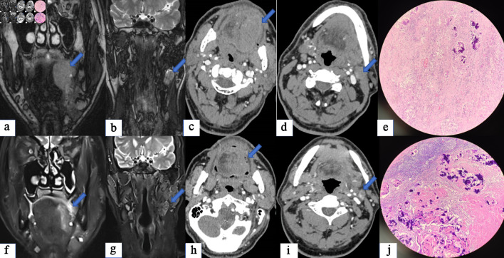
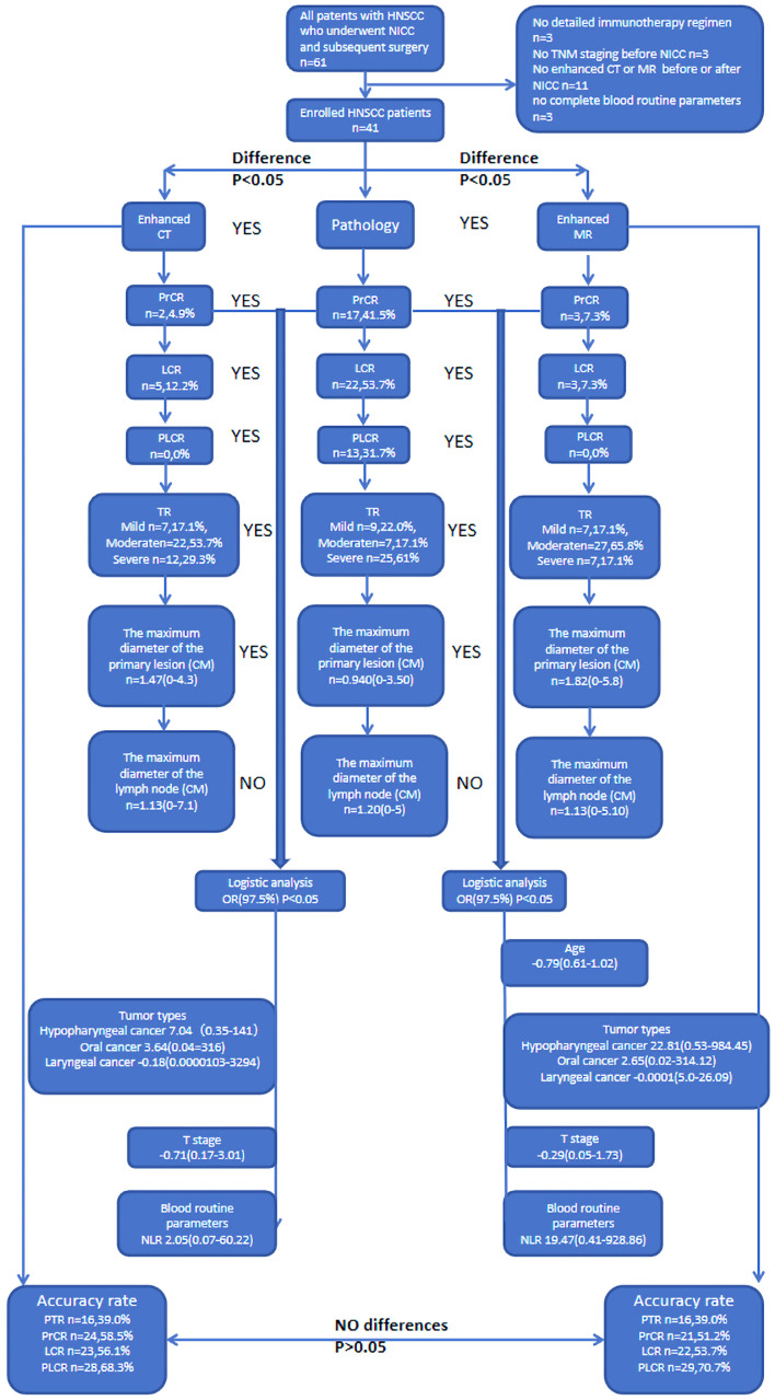
Methods: HNSCC patients who underwent NICC and surgery from May 2021 to September 2023 were retrospectively analyzed. All patients had to undergo imaging examination evaluation, including enhanced computed tomography (CT) and enhanced magnetic resonance (MR) imaging before and after NICC. Data related to clinical parameters, complete response of the primary site (PrCR), complete response of the primary site and the lymph node (PLCR), complete response of the lymph node (LCR), and tumor response (TR), were gathered. The paired Chi-square test and t-test were conducted to analyze the differences in responses between imaging examination and pathology. Binary logistic regression was applied to analyze the relevant clinical factors of differences in responses.
Results: In total, data of 41 patients were included in this study. Significant discordant responses were observed between enhanced CT, magnetic resonance imaging (MRI), and pathology in PrCR (4.9%, 7.3% vs. 41.5%), LCR (12.2%, 7.3% vs. 53.7%), PLCR (0%, 0% vs. 31.7%), and TR (severe 29.3%,17.1% vs. 25.61%) (P < 0.05). Patients with hypopharyngeal cancer (odds ratio (OR): 7.04), oral cancer (OR: 3.64), higher neutrophil to lymphocyte ratio (NLR) (OR: 2.05), and earlier T stage (OR: 0.71) exhibited a larger response difference between enhanced CT and pathology. Patients with younger age (OR: 0.79) hypopharyngeal cancer (OR: 22.81), oral cancer (OR: 2.65), higher NLR (OR: 19.47), and earlier T stage (OR: 0.29) exhibited a larger response difference between enhanced MR and pathology.
Conclusions: Discordant responses were noted between the imaging examination and surgical pathology of HNSCC after NICC. Hypopharyngeal cancer, higher NLR, and earlier T stage may predict a higher response difference.
期刊介绍:
World Journal of Oncology, bimonthly, publishes original contributions describing basic research and clinical investigation of cancer, on the cellular, molecular, prevention, diagnosis, therapy and prognosis aspects. The submissions can be basic research or clinical investigation oriented. This journal welcomes those submissions focused on the clinical trials of new treatment modalities for cancer, and those submissions focused on molecular or cellular research of the oncology pathogenesis. Case reports submitted for consideration of publication should explore either a novel genomic event/description or a new safety signal from an oncolytic agent. The areas of interested manuscripts are these disciplines: tumor immunology and immunotherapy; cancer molecular pharmacology and chemotherapy; drug sensitivity and resistance; cancer epidemiology; clinical trials; cancer pathology; radiobiology and radiation oncology; solid tumor oncology; hematological malignancies; surgical oncology; pediatric oncology; molecular oncology and cancer genes; gene therapy; cancer endocrinology; cancer metastasis; prevention and diagnosis of cancer; other cancer related subjects. The types of manuscripts accepted are original article, review, editorial, short communication, case report, letter to the editor, book review.

 求助内容:
求助内容: 应助结果提醒方式:


