Maxime Ablefoni, Theresa Richter, Jakob Leonhardi, Constantin Ehrengut, Gordian Prasse, Matthias Mehdorn, Daniel Seehofer, Anne Kathrin Höhn, Timm Denecke, Hans-Jonas Meyer
{"title":"高b值计算机扩散加权成像对肝细胞癌的潜在诊断价值。","authors":"Maxime Ablefoni, Theresa Richter, Jakob Leonhardi, Constantin Ehrengut, Gordian Prasse, Matthias Mehdorn, Daniel Seehofer, Anne Kathrin Höhn, Timm Denecke, Hans-Jonas Meyer","doi":"10.5114/ceh.2024.139651","DOIUrl":null,"url":null,"abstract":"<p><strong>Aim of the study: </strong>Over the past few years, diffusion-weighted imaging (DWI) has become an increasingly important diagnostic tool in the diagnosis of liver lesions. The objective of the present study was to evaluate the diagnostic benefit of high b-value computed diffusion-weighted imaging (c-DWI) compared with standard DWI in patients with hepatocellular carcinoma (HCC) and whether there is an association with microvascular invasion (MVI).</p><p><strong>Material and methods: </strong>In total, 37 patients with histopathologically confirmed HCC were retrospectively ana-lyzed. DWI was acquired with b-values of 50, 400, and 800 or 1000 s/mm² on a 1.5 T magnetic resonance imaging (MRI) scanner. The c-DWI was calculated using a monoexponential model with high b-values of 1000, 2000, 3000, 4000, and 5000 s/mm². All high b-value c-DWI images were compared to the standard DWI in terms of volume, detectability of hepatic lesions, and image quality.</p><p><strong>Results: </strong>Regarding lesion volume and image quality there were no statistically significant differences between standard and c-DWI. HCC lesions measured on DWI images were statistically significantly larger compared to c-DWI images starting from a b value of 2000 s/mm<sup>2</sup> (DWI vs. c-DWI b 2000 s/mm<sup>2</sup>: 2 cm<sup>3</sup> [1-12] cm<sup>3</sup> vs. 1 cm<sup>3</sup> [0-17] cm<sup>3</sup>, <i>p</i> < 0.05). Moreover, there was deterioration of image quality starting at b = 2000 s/mm<sup>2</sup>. There were no significant differences in terms of lesion signal intensity in DWI and c-DWI images. There were no differences for the DWI parameters according to MVI status.</p><p><strong>Conclusions: </strong>C-DWI images with high b-values up to b = 1000 s/mm<sup>2</sup> demonstrate comparable detectability of HCC compared to standard DWI. The investigated DWI parameters were not associated with MVI status. Further research is needed to evaluate the potential benefit of high b-value c-DWI.</p>","PeriodicalId":10281,"journal":{"name":"Clinical and Experimental Hepatology","volume":"10 2","pages":"129-136"},"PeriodicalIF":1.7000,"publicationDate":"2024-03-01","publicationTypes":"Journal Article","fieldsOfStudy":null,"isOpenAccess":false,"openAccessPdf":"https://www.ncbi.nlm.nih.gov/pmc/articles/PMC11748228/pdf/","citationCount":"0","resultStr":"{\"title\":\"Potential diagnostic value of high b-value computed diffusion-weighted imaging in hepatocellular carcinoma.\",\"authors\":\"Maxime Ablefoni, Theresa Richter, Jakob Leonhardi, Constantin Ehrengut, Gordian Prasse, Matthias Mehdorn, Daniel Seehofer, Anne Kathrin Höhn, Timm Denecke, Hans-Jonas Meyer\",\"doi\":\"10.5114/ceh.2024.139651\",\"DOIUrl\":null,\"url\":null,\"abstract\":\"<p><strong>Aim of the study: </strong>Over the past few years, diffusion-weighted imaging (DWI) has become an increasingly important diagnostic tool in the diagnosis of liver lesions. The objective of the present study was to evaluate the diagnostic benefit of high b-value computed diffusion-weighted imaging (c-DWI) compared with standard DWI in patients with hepatocellular carcinoma (HCC) and whether there is an association with microvascular invasion (MVI).</p><p><strong>Material and methods: </strong>In total, 37 patients with histopathologically confirmed HCC were retrospectively ana-lyzed. DWI was acquired with b-values of 50, 400, and 800 or 1000 s/mm² on a 1.5 T magnetic resonance imaging (MRI) scanner. The c-DWI was calculated using a monoexponential model with high b-values of 1000, 2000, 3000, 4000, and 5000 s/mm². All high b-value c-DWI images were compared to the standard DWI in terms of volume, detectability of hepatic lesions, and image quality.</p><p><strong>Results: </strong>Regarding lesion volume and image quality there were no statistically significant differences between standard and c-DWI. HCC lesions measured on DWI images were statistically significantly larger compared to c-DWI images starting from a b value of 2000 s/mm<sup>2</sup> (DWI vs. c-DWI b 2000 s/mm<sup>2</sup>: 2 cm<sup>3</sup> [1-12] cm<sup>3</sup> vs. 1 cm<sup>3</sup> [0-17] cm<sup>3</sup>, <i>p</i> < 0.05). Moreover, there was deterioration of image quality starting at b = 2000 s/mm<sup>2</sup>. There were no significant differences in terms of lesion signal intensity in DWI and c-DWI images. There were no differences for the DWI parameters according to MVI status.</p><p><strong>Conclusions: </strong>C-DWI images with high b-values up to b = 1000 s/mm<sup>2</sup> demonstrate comparable detectability of HCC compared to standard DWI. The investigated DWI parameters were not associated with MVI status. Further research is needed to evaluate the potential benefit of high b-value c-DWI.</p>\",\"PeriodicalId\":10281,\"journal\":{\"name\":\"Clinical and Experimental Hepatology\",\"volume\":\"10 2\",\"pages\":\"129-136\"},\"PeriodicalIF\":1.7000,\"publicationDate\":\"2024-03-01\",\"publicationTypes\":\"Journal Article\",\"fieldsOfStudy\":null,\"isOpenAccess\":false,\"openAccessPdf\":\"https://www.ncbi.nlm.nih.gov/pmc/articles/PMC11748228/pdf/\",\"citationCount\":\"0\",\"resultStr\":null,\"platform\":\"Semanticscholar\",\"paperid\":null,\"PeriodicalName\":\"Clinical and Experimental Hepatology\",\"FirstCategoryId\":\"1085\",\"ListUrlMain\":\"https://doi.org/10.5114/ceh.2024.139651\",\"RegionNum\":0,\"RegionCategory\":null,\"ArticlePicture\":[],\"TitleCN\":null,\"AbstractTextCN\":null,\"PMCID\":null,\"EPubDate\":\"2024/6/11 0:00:00\",\"PubModel\":\"Epub\",\"JCR\":\"Q3\",\"JCRName\":\"GASTROENTEROLOGY & HEPATOLOGY\",\"Score\":null,\"Total\":0}","platform":"Semanticscholar","paperid":null,"PeriodicalName":"Clinical and Experimental Hepatology","FirstCategoryId":"1085","ListUrlMain":"https://doi.org/10.5114/ceh.2024.139651","RegionNum":0,"RegionCategory":null,"ArticlePicture":[],"TitleCN":null,"AbstractTextCN":null,"PMCID":null,"EPubDate":"2024/6/11 0:00:00","PubModel":"Epub","JCR":"Q3","JCRName":"GASTROENTEROLOGY & HEPATOLOGY","Score":null,"Total":0}
引用次数: 0
摘要
研究目的:近年来,弥散加权成像(DWI)已成为肝脏病变诊断中越来越重要的诊断工具。本研究的目的是评估高b值计算机弥散加权成像(c-DWI)与标准DWI在肝细胞癌(HCC)患者中的诊断价值,以及是否与微血管侵袭(MVI)相关。材料与方法:对37例经组织病理学证实的HCC患者进行回顾性分析。在1.5 T磁共振成像(MRI)扫描仪上获得b值为50、400、800或1000 s/mm²的DWI。c-DWI采用单指数模型计算,高b值为1000、2000、3000、4000和5000 s/mm²。将所有高b值c-DWI图像与标准DWI在体积、肝脏病变的可检测性和图像质量方面进行比较。结果:在病变体积和图像质量方面,标准与c-DWI差异无统计学意义。从b值2000 s/mm2开始,DWI图像上测量的HCC病变与c-DWI图像相比,有统计学意义上更大(DWI vs c-DWI b 2000 s/mm2: 2 cm3 [1-12] cm3 vs 1 cm3 [0-17] cm3, p < 0.05)。此外,从b = 2000 s/mm2开始,图像质量开始恶化。DWI和c-DWI图像的病变信号强度差异无统计学意义。不同MVI状态的DWI参数无差异。结论:与标准DWI相比,高b值高达b = 1000 s/mm2的C-DWI图像具有相当的HCC检出率。所调查的DWI参数与MVI状态无关。高b值c-DWI的潜在益处有待进一步研究。
Potential diagnostic value of high b-value computed diffusion-weighted imaging in hepatocellular carcinoma.
Aim of the study: Over the past few years, diffusion-weighted imaging (DWI) has become an increasingly important diagnostic tool in the diagnosis of liver lesions. The objective of the present study was to evaluate the diagnostic benefit of high b-value computed diffusion-weighted imaging (c-DWI) compared with standard DWI in patients with hepatocellular carcinoma (HCC) and whether there is an association with microvascular invasion (MVI).
Material and methods: In total, 37 patients with histopathologically confirmed HCC were retrospectively ana-lyzed. DWI was acquired with b-values of 50, 400, and 800 or 1000 s/mm² on a 1.5 T magnetic resonance imaging (MRI) scanner. The c-DWI was calculated using a monoexponential model with high b-values of 1000, 2000, 3000, 4000, and 5000 s/mm². All high b-value c-DWI images were compared to the standard DWI in terms of volume, detectability of hepatic lesions, and image quality.
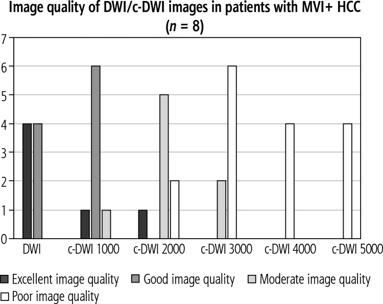
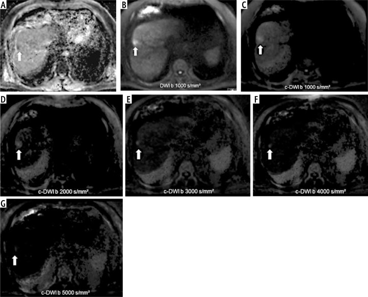
Results: Regarding lesion volume and image quality there were no statistically significant differences between standard and c-DWI. HCC lesions measured on DWI images were statistically significantly larger compared to c-DWI images starting from a b value of 2000 s/mm2 (DWI vs. c-DWI b 2000 s/mm2: 2 cm3 [1-12] cm3 vs. 1 cm3 [0-17] cm3, p < 0.05). Moreover, there was deterioration of image quality starting at b = 2000 s/mm2. There were no significant differences in terms of lesion signal intensity in DWI and c-DWI images. There were no differences for the DWI parameters according to MVI status.
Conclusions: C-DWI images with high b-values up to b = 1000 s/mm2 demonstrate comparable detectability of HCC compared to standard DWI. The investigated DWI parameters were not associated with MVI status. Further research is needed to evaluate the potential benefit of high b-value c-DWI.
期刊介绍:
Clinical and Experimental Hepatology – quarterly of the Polish Association for Study of Liver – is a scientific and educational, peer-reviewed journal publishing original and review papers describing clinical and basic investigations in the field of hepatology.

 求助内容:
求助内容: 应助结果提醒方式:


