{"title":"吲哚菁绿粪便具有诊断新生儿胆道闭锁的潜力。","authors":"Mika Murayama, Toshihiro Yasui, Mikihiro Inoue, Shunsuke Watanabe, Atsuki Naoe, Yasuhiro Kondo, Tomonori Tsuchiya, Tatsuya Suzuki","doi":"10.5114/ceh.2024.139979","DOIUrl":null,"url":null,"abstract":"<p><strong>Aim of the study: </strong>This study aimed to establish an objective, simple, and minimally invasive screening method to detect patients with biliary atresia during neonatal checkups by using indocyanine green (ICG) fluorescence in the stool.</p><p><strong>Material and methods: </strong>We produced a rat model of extrahepatic biliary obstruction (group O, <i>n</i> = 9) and compared the stools from these rats with those of control group rats (group C, <i>n</i> = 6) by a fluorescence technique. ICG was administered (0.5 mg/kg) through the caudal vein; group O received ICG at the end of surgery.</p><p><strong>Results: </strong>In group C, we collected stools at 3, 6, 12, 24, 48, and 72 hours, and fluorescence disappeared at 48 hours. In group O, stools were collected at 24, 48, 72, 96, and 120 hours after surgery, and fluorescence continued at 120 hours without the loss of fluorescence. Quantitative assessment of lightness showed significant differences between the groups at 48 and 72 hours (<i>p</i> = 0.0016 and <i>p</i> = 0.0004, respectively).</p><p><strong>Conclusions: </strong>This study shows that ICG is excreted into the gastrointestinal tract via a route other than the bile duct in a rat model of extrahepatic biliary obstruction. Our findings also suggest that ICG has the potential for initial screening of biliary congestive disease in the neonatal period, which could be followed up by detailed testing.</p>","PeriodicalId":10281,"journal":{"name":"Clinical and Experimental Hepatology","volume":"10 2","pages":"98-103"},"PeriodicalIF":1.7000,"publicationDate":"2024-03-01","publicationTypes":"Journal Article","fieldsOfStudy":null,"isOpenAccess":false,"openAccessPdf":"https://www.ncbi.nlm.nih.gov/pmc/articles/PMC11748229/pdf/","citationCount":"0","resultStr":"{\"title\":\"Indocyanine green faecal excretion holds potential for diagnosis of neonatal biliary atresia.\",\"authors\":\"Mika Murayama, Toshihiro Yasui, Mikihiro Inoue, Shunsuke Watanabe, Atsuki Naoe, Yasuhiro Kondo, Tomonori Tsuchiya, Tatsuya Suzuki\",\"doi\":\"10.5114/ceh.2024.139979\",\"DOIUrl\":null,\"url\":null,\"abstract\":\"<p><strong>Aim of the study: </strong>This study aimed to establish an objective, simple, and minimally invasive screening method to detect patients with biliary atresia during neonatal checkups by using indocyanine green (ICG) fluorescence in the stool.</p><p><strong>Material and methods: </strong>We produced a rat model of extrahepatic biliary obstruction (group O, <i>n</i> = 9) and compared the stools from these rats with those of control group rats (group C, <i>n</i> = 6) by a fluorescence technique. ICG was administered (0.5 mg/kg) through the caudal vein; group O received ICG at the end of surgery.</p><p><strong>Results: </strong>In group C, we collected stools at 3, 6, 12, 24, 48, and 72 hours, and fluorescence disappeared at 48 hours. In group O, stools were collected at 24, 48, 72, 96, and 120 hours after surgery, and fluorescence continued at 120 hours without the loss of fluorescence. Quantitative assessment of lightness showed significant differences between the groups at 48 and 72 hours (<i>p</i> = 0.0016 and <i>p</i> = 0.0004, respectively).</p><p><strong>Conclusions: </strong>This study shows that ICG is excreted into the gastrointestinal tract via a route other than the bile duct in a rat model of extrahepatic biliary obstruction. Our findings also suggest that ICG has the potential for initial screening of biliary congestive disease in the neonatal period, which could be followed up by detailed testing.</p>\",\"PeriodicalId\":10281,\"journal\":{\"name\":\"Clinical and Experimental Hepatology\",\"volume\":\"10 2\",\"pages\":\"98-103\"},\"PeriodicalIF\":1.7000,\"publicationDate\":\"2024-03-01\",\"publicationTypes\":\"Journal Article\",\"fieldsOfStudy\":null,\"isOpenAccess\":false,\"openAccessPdf\":\"https://www.ncbi.nlm.nih.gov/pmc/articles/PMC11748229/pdf/\",\"citationCount\":\"0\",\"resultStr\":null,\"platform\":\"Semanticscholar\",\"paperid\":null,\"PeriodicalName\":\"Clinical and Experimental Hepatology\",\"FirstCategoryId\":\"1085\",\"ListUrlMain\":\"https://doi.org/10.5114/ceh.2024.139979\",\"RegionNum\":0,\"RegionCategory\":null,\"ArticlePicture\":[],\"TitleCN\":null,\"AbstractTextCN\":null,\"PMCID\":null,\"EPubDate\":\"2024/5/29 0:00:00\",\"PubModel\":\"Epub\",\"JCR\":\"Q3\",\"JCRName\":\"GASTROENTEROLOGY & HEPATOLOGY\",\"Score\":null,\"Total\":0}","platform":"Semanticscholar","paperid":null,"PeriodicalName":"Clinical and Experimental Hepatology","FirstCategoryId":"1085","ListUrlMain":"https://doi.org/10.5114/ceh.2024.139979","RegionNum":0,"RegionCategory":null,"ArticlePicture":[],"TitleCN":null,"AbstractTextCN":null,"PMCID":null,"EPubDate":"2024/5/29 0:00:00","PubModel":"Epub","JCR":"Q3","JCRName":"GASTROENTEROLOGY & HEPATOLOGY","Score":null,"Total":0}
Indocyanine green faecal excretion holds potential for diagnosis of neonatal biliary atresia.
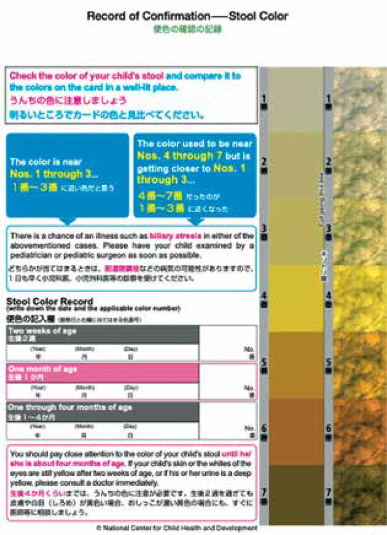
Aim of the study: This study aimed to establish an objective, simple, and minimally invasive screening method to detect patients with biliary atresia during neonatal checkups by using indocyanine green (ICG) fluorescence in the stool.
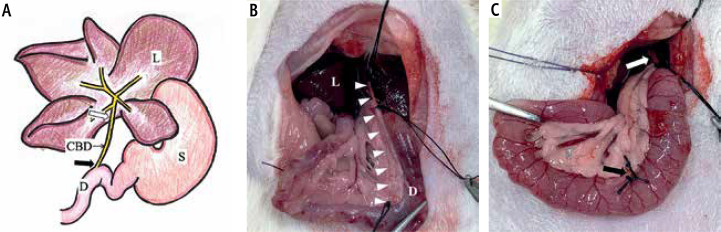
Material and methods: We produced a rat model of extrahepatic biliary obstruction (group O, n = 9) and compared the stools from these rats with those of control group rats (group C, n = 6) by a fluorescence technique. ICG was administered (0.5 mg/kg) through the caudal vein; group O received ICG at the end of surgery.
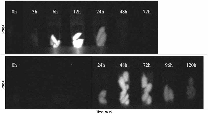
Results: In group C, we collected stools at 3, 6, 12, 24, 48, and 72 hours, and fluorescence disappeared at 48 hours. In group O, stools were collected at 24, 48, 72, 96, and 120 hours after surgery, and fluorescence continued at 120 hours without the loss of fluorescence. Quantitative assessment of lightness showed significant differences between the groups at 48 and 72 hours (p = 0.0016 and p = 0.0004, respectively).
Conclusions: This study shows that ICG is excreted into the gastrointestinal tract via a route other than the bile duct in a rat model of extrahepatic biliary obstruction. Our findings also suggest that ICG has the potential for initial screening of biliary congestive disease in the neonatal period, which could be followed up by detailed testing.
期刊介绍:
Clinical and Experimental Hepatology – quarterly of the Polish Association for Study of Liver – is a scientific and educational, peer-reviewed journal publishing original and review papers describing clinical and basic investigations in the field of hepatology.

 求助内容:
求助内容: 应助结果提醒方式:


