{"title":"神经母细胞瘤高危组与非高危组解剖影像学特征的分层。","authors":"Haoru Wang, Xin Chen, Ling He, Jinhua Cai","doi":"10.1177/10732748251315883","DOIUrl":null,"url":null,"abstract":"<p><strong>Background: </strong>This study compared anatomical imaging features between high-risk and non-high-risk groups in neuroblastoma with at least one image-defined risk factor (IDRF). It also assessed the diagnostic performance of these features in identifying the high-risk group.</p><p><strong>Methods: </strong>A retrospective analysis of neuroblastoma patients with at least one IDRF was conducted. Imaging features, including estimated tumor volume and IDRFs, were compared between the two groups. The diagnostic performance of these features was assessed using receiver operating characteristic (ROC) curves, and the areas under the ROC curves (AUCs) along with their 95% confidence intervals (CIs) were calculated. Additionally, to internally validate their diagnostic performance, the bootstrap resampling method with 1000 bootstrap resamples was employed.</p><p><strong>Results: </strong>The study included 255 patients (185 high-risk cases, 70 non-high-risk cases). Significant differences were found in estimated tumor volume and IDRF number between the high-risk and non-high-risk groups (<i>P</i> < 0.001). The estimated tumor volume and the IDRF number-based cluster were independent risk factors, and their combination achieved an AUC of 0.801 (95% CI: 0.747-0.848) for high-risk group diagnosis, with the average AUC of the 1000 bootstrap samples of 0.800 (95% CI: 0.798-0.802). In abdominal lesions, specific IDRF categories differed between high-risk and non-high-risk groups (<i>P</i> < 0.05).</p><p><strong>Conclusion: </strong>Our study reveals anatomical imaging differences between high-risk and non-high-risk groups in neuroblastoma with at least one IDRF.</p>","PeriodicalId":49093,"journal":{"name":"Cancer Control","volume":"32 ","pages":"10732748251315883"},"PeriodicalIF":2.6000,"publicationDate":"2025-01-01","publicationTypes":"Journal Article","fieldsOfStudy":null,"isOpenAccess":false,"openAccessPdf":"https://www.ncbi.nlm.nih.gov/pmc/articles/PMC11748161/pdf/","citationCount":"0","resultStr":"{\"title\":\"Stratification of Anatomical Imaging Features Between High-Risk and Non-High-Risk Groups in Neuroblastoma.\",\"authors\":\"Haoru Wang, Xin Chen, Ling He, Jinhua Cai\",\"doi\":\"10.1177/10732748251315883\",\"DOIUrl\":null,\"url\":null,\"abstract\":\"<p><strong>Background: </strong>This study compared anatomical imaging features between high-risk and non-high-risk groups in neuroblastoma with at least one image-defined risk factor (IDRF). It also assessed the diagnostic performance of these features in identifying the high-risk group.</p><p><strong>Methods: </strong>A retrospective analysis of neuroblastoma patients with at least one IDRF was conducted. Imaging features, including estimated tumor volume and IDRFs, were compared between the two groups. The diagnostic performance of these features was assessed using receiver operating characteristic (ROC) curves, and the areas under the ROC curves (AUCs) along with their 95% confidence intervals (CIs) were calculated. Additionally, to internally validate their diagnostic performance, the bootstrap resampling method with 1000 bootstrap resamples was employed.</p><p><strong>Results: </strong>The study included 255 patients (185 high-risk cases, 70 non-high-risk cases). Significant differences were found in estimated tumor volume and IDRF number between the high-risk and non-high-risk groups (<i>P</i> < 0.001). The estimated tumor volume and the IDRF number-based cluster were independent risk factors, and their combination achieved an AUC of 0.801 (95% CI: 0.747-0.848) for high-risk group diagnosis, with the average AUC of the 1000 bootstrap samples of 0.800 (95% CI: 0.798-0.802). In abdominal lesions, specific IDRF categories differed between high-risk and non-high-risk groups (<i>P</i> < 0.05).</p><p><strong>Conclusion: </strong>Our study reveals anatomical imaging differences between high-risk and non-high-risk groups in neuroblastoma with at least one IDRF.</p>\",\"PeriodicalId\":49093,\"journal\":{\"name\":\"Cancer Control\",\"volume\":\"32 \",\"pages\":\"10732748251315883\"},\"PeriodicalIF\":2.6000,\"publicationDate\":\"2025-01-01\",\"publicationTypes\":\"Journal Article\",\"fieldsOfStudy\":null,\"isOpenAccess\":false,\"openAccessPdf\":\"https://www.ncbi.nlm.nih.gov/pmc/articles/PMC11748161/pdf/\",\"citationCount\":\"0\",\"resultStr\":null,\"platform\":\"Semanticscholar\",\"paperid\":null,\"PeriodicalName\":\"Cancer Control\",\"FirstCategoryId\":\"3\",\"ListUrlMain\":\"https://doi.org/10.1177/10732748251315883\",\"RegionNum\":4,\"RegionCategory\":\"医学\",\"ArticlePicture\":[],\"TitleCN\":null,\"AbstractTextCN\":null,\"PMCID\":null,\"EPubDate\":\"\",\"PubModel\":\"\",\"JCR\":\"Q3\",\"JCRName\":\"ONCOLOGY\",\"Score\":null,\"Total\":0}","platform":"Semanticscholar","paperid":null,"PeriodicalName":"Cancer Control","FirstCategoryId":"3","ListUrlMain":"https://doi.org/10.1177/10732748251315883","RegionNum":4,"RegionCategory":"医学","ArticlePicture":[],"TitleCN":null,"AbstractTextCN":null,"PMCID":null,"EPubDate":"","PubModel":"","JCR":"Q3","JCRName":"ONCOLOGY","Score":null,"Total":0}
Stratification of Anatomical Imaging Features Between High-Risk and Non-High-Risk Groups in Neuroblastoma.
Background: This study compared anatomical imaging features between high-risk and non-high-risk groups in neuroblastoma with at least one image-defined risk factor (IDRF). It also assessed the diagnostic performance of these features in identifying the high-risk group.
Methods: A retrospective analysis of neuroblastoma patients with at least one IDRF was conducted. Imaging features, including estimated tumor volume and IDRFs, were compared between the two groups. The diagnostic performance of these features was assessed using receiver operating characteristic (ROC) curves, and the areas under the ROC curves (AUCs) along with their 95% confidence intervals (CIs) were calculated. Additionally, to internally validate their diagnostic performance, the bootstrap resampling method with 1000 bootstrap resamples was employed.
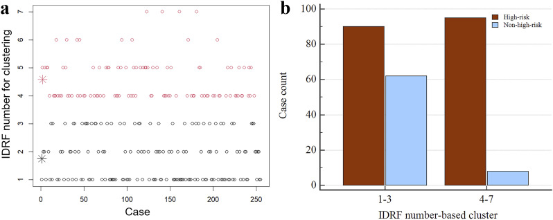
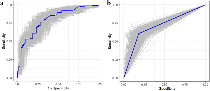
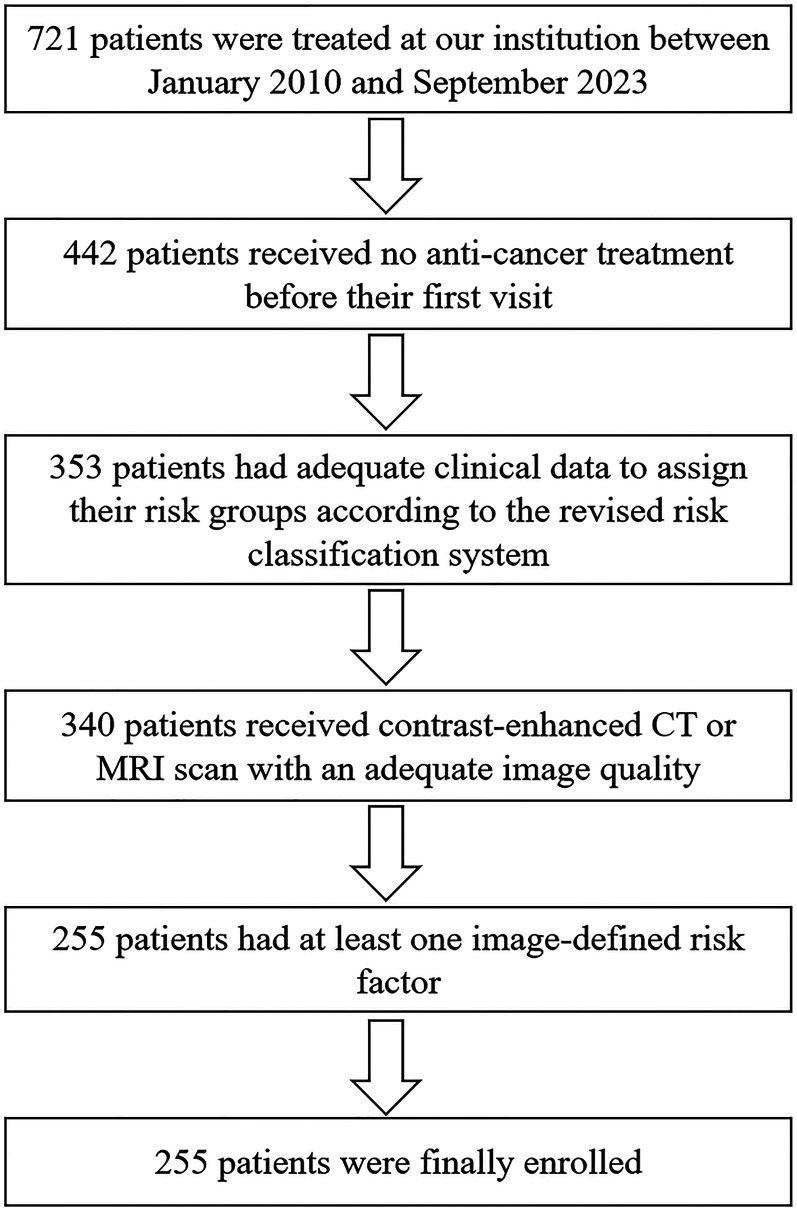
Results: The study included 255 patients (185 high-risk cases, 70 non-high-risk cases). Significant differences were found in estimated tumor volume and IDRF number between the high-risk and non-high-risk groups (P < 0.001). The estimated tumor volume and the IDRF number-based cluster were independent risk factors, and their combination achieved an AUC of 0.801 (95% CI: 0.747-0.848) for high-risk group diagnosis, with the average AUC of the 1000 bootstrap samples of 0.800 (95% CI: 0.798-0.802). In abdominal lesions, specific IDRF categories differed between high-risk and non-high-risk groups (P < 0.05).
Conclusion: Our study reveals anatomical imaging differences between high-risk and non-high-risk groups in neuroblastoma with at least one IDRF.
期刊介绍:
Cancer Control is a JCR-ranked, peer-reviewed open access journal whose mission is to advance the prevention, detection, diagnosis, treatment, and palliative care of cancer by enabling researchers, doctors, policymakers, and other healthcare professionals to freely share research along the cancer control continuum. Our vision is a world where gold-standard cancer care is the norm, not the exception.
 求助内容:
求助内容: 应助结果提醒方式:


