Anouar Bourghli, Yaser Almonla, Louis Boissiere, Faisal Konbaz, Khaled Almusrea, Ibrahim Obeid
{"title":"弥漫性特发性骨骼增生引起的吞咽困难的外科治疗:钡餐透视的作用- 1例报告。","authors":"Anouar Bourghli, Yaser Almonla, Louis Boissiere, Faisal Konbaz, Khaled Almusrea, Ibrahim Obeid","doi":"10.21037/jss-24-84","DOIUrl":null,"url":null,"abstract":"<p><strong>Background: </strong>Diffuse idiopathic skeletal hyperostosis (DISH) is a systemic condition that might lead to dysphagia because of massive osteophytes that may be present at multiple levels. Confirming the symptomatic level to guide surgical management and avoid extensive surgery is important, however, there is no globally accepted consensus on the topic.</p><p><strong>Case description: </strong>We report the case of a 51-year-old man, with no specific past medical history, who has been complaining of a 3-months pain in the left side of the tongue base with sensation of a lump in the throat and dysphagia. Computed tomography scan confirmed DISH between C4 and C7. Barium swallow fluoroscopy demonstrated indentation of the esophagus only at the level of C4-C5, which guided the surgical management that focused on resecting only the major osteophytes at the level of C4-C5, avoiding extensive approach with its subsequent possible complications, and enabling satisfactory clinical and radiological outcomes.</p><p><strong>Conclusions: </strong>The current case thoroughly illustrated the diagnosis and surgical management in the presence of dysphagia from DISH. Through an anterior pre-vascular approach complete resection of the major osteophytes could be done. Barium swallow fluoroscopy showed very high interest in dynamically demonstrating the main level causing the dysphagia symptoms and also confirming satisfactory esophagus decompression and release after surgery.</p>","PeriodicalId":17131,"journal":{"name":"Journal of spine surgery","volume":"10 4","pages":"758-763"},"PeriodicalIF":0.0000,"publicationDate":"2024-12-20","publicationTypes":"Journal Article","fieldsOfStudy":null,"isOpenAccess":false,"openAccessPdf":"https://www.ncbi.nlm.nih.gov/pmc/articles/PMC11732326/pdf/","citationCount":"0","resultStr":"{\"title\":\"Surgical management of dysphagia due to diffuse idiopathic skeletal hyperostosis: the role of barium swallow fluoroscopy-a case report.\",\"authors\":\"Anouar Bourghli, Yaser Almonla, Louis Boissiere, Faisal Konbaz, Khaled Almusrea, Ibrahim Obeid\",\"doi\":\"10.21037/jss-24-84\",\"DOIUrl\":null,\"url\":null,\"abstract\":\"<p><strong>Background: </strong>Diffuse idiopathic skeletal hyperostosis (DISH) is a systemic condition that might lead to dysphagia because of massive osteophytes that may be present at multiple levels. Confirming the symptomatic level to guide surgical management and avoid extensive surgery is important, however, there is no globally accepted consensus on the topic.</p><p><strong>Case description: </strong>We report the case of a 51-year-old man, with no specific past medical history, who has been complaining of a 3-months pain in the left side of the tongue base with sensation of a lump in the throat and dysphagia. Computed tomography scan confirmed DISH between C4 and C7. Barium swallow fluoroscopy demonstrated indentation of the esophagus only at the level of C4-C5, which guided the surgical management that focused on resecting only the major osteophytes at the level of C4-C5, avoiding extensive approach with its subsequent possible complications, and enabling satisfactory clinical and radiological outcomes.</p><p><strong>Conclusions: </strong>The current case thoroughly illustrated the diagnosis and surgical management in the presence of dysphagia from DISH. Through an anterior pre-vascular approach complete resection of the major osteophytes could be done. Barium swallow fluoroscopy showed very high interest in dynamically demonstrating the main level causing the dysphagia symptoms and also confirming satisfactory esophagus decompression and release after surgery.</p>\",\"PeriodicalId\":17131,\"journal\":{\"name\":\"Journal of spine surgery\",\"volume\":\"10 4\",\"pages\":\"758-763\"},\"PeriodicalIF\":0.0000,\"publicationDate\":\"2024-12-20\",\"publicationTypes\":\"Journal Article\",\"fieldsOfStudy\":null,\"isOpenAccess\":false,\"openAccessPdf\":\"https://www.ncbi.nlm.nih.gov/pmc/articles/PMC11732326/pdf/\",\"citationCount\":\"0\",\"resultStr\":null,\"platform\":\"Semanticscholar\",\"paperid\":null,\"PeriodicalName\":\"Journal of spine surgery\",\"FirstCategoryId\":\"1085\",\"ListUrlMain\":\"https://doi.org/10.21037/jss-24-84\",\"RegionNum\":0,\"RegionCategory\":null,\"ArticlePicture\":[],\"TitleCN\":null,\"AbstractTextCN\":null,\"PMCID\":null,\"EPubDate\":\"2024/12/11 0:00:00\",\"PubModel\":\"Epub\",\"JCR\":\"Q1\",\"JCRName\":\"Medicine\",\"Score\":null,\"Total\":0}","platform":"Semanticscholar","paperid":null,"PeriodicalName":"Journal of spine surgery","FirstCategoryId":"1085","ListUrlMain":"https://doi.org/10.21037/jss-24-84","RegionNum":0,"RegionCategory":null,"ArticlePicture":[],"TitleCN":null,"AbstractTextCN":null,"PMCID":null,"EPubDate":"2024/12/11 0:00:00","PubModel":"Epub","JCR":"Q1","JCRName":"Medicine","Score":null,"Total":0}
Surgical management of dysphagia due to diffuse idiopathic skeletal hyperostosis: the role of barium swallow fluoroscopy-a case report.
Background: Diffuse idiopathic skeletal hyperostosis (DISH) is a systemic condition that might lead to dysphagia because of massive osteophytes that may be present at multiple levels. Confirming the symptomatic level to guide surgical management and avoid extensive surgery is important, however, there is no globally accepted consensus on the topic.
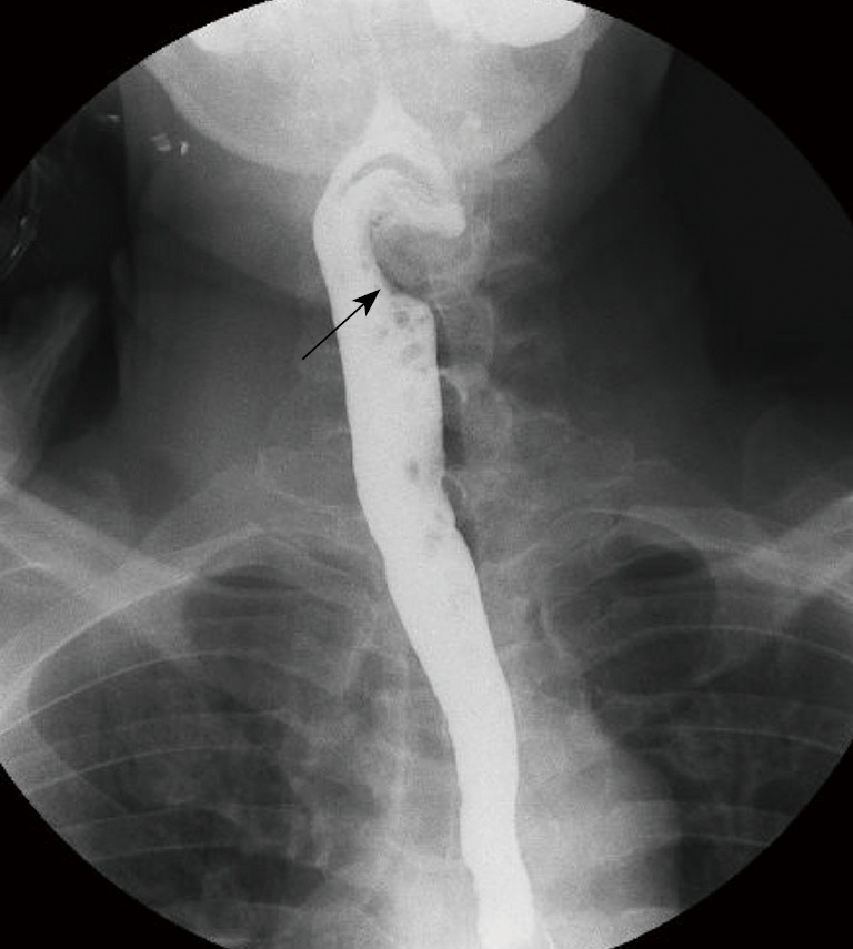
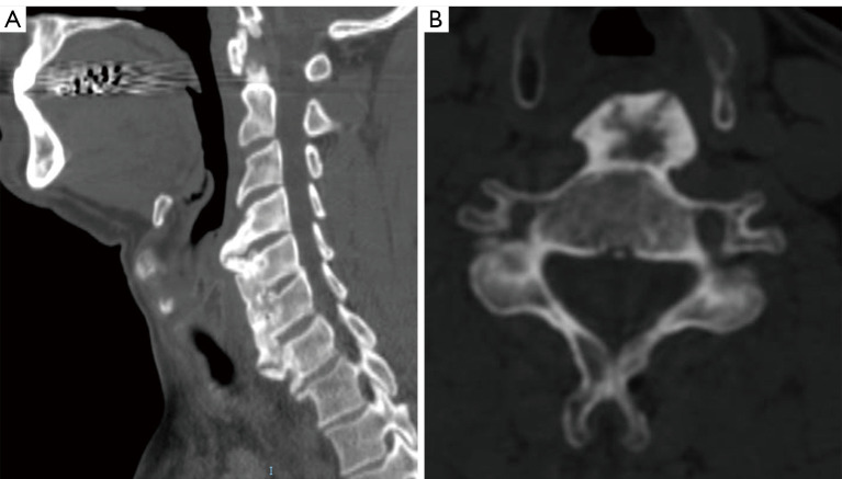
Case description: We report the case of a 51-year-old man, with no specific past medical history, who has been complaining of a 3-months pain in the left side of the tongue base with sensation of a lump in the throat and dysphagia. Computed tomography scan confirmed DISH between C4 and C7. Barium swallow fluoroscopy demonstrated indentation of the esophagus only at the level of C4-C5, which guided the surgical management that focused on resecting only the major osteophytes at the level of C4-C5, avoiding extensive approach with its subsequent possible complications, and enabling satisfactory clinical and radiological outcomes.
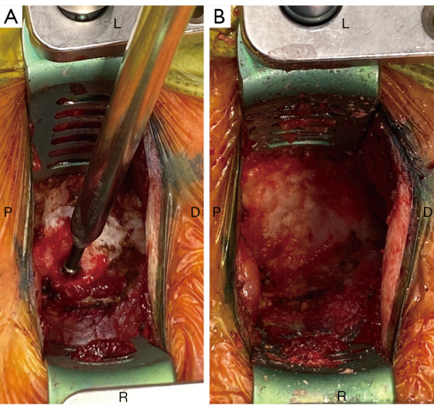
Conclusions: The current case thoroughly illustrated the diagnosis and surgical management in the presence of dysphagia from DISH. Through an anterior pre-vascular approach complete resection of the major osteophytes could be done. Barium swallow fluoroscopy showed very high interest in dynamically demonstrating the main level causing the dysphagia symptoms and also confirming satisfactory esophagus decompression and release after surgery.

 求助内容:
求助内容: 应助结果提醒方式:


