Soumaya Debiche, Houda Snene, Bochra Bouchabou, Monia Attia, Hana Blibech, Aïda Ayadi, Rym Enneifer, Leila Ben Farhat, Nadia Mehiri, Bechir Louzir
{"title":"胸外肿瘤支气管内转移的诊断与治疗方法:附三例报告及文献复习。","authors":"Soumaya Debiche, Houda Snene, Bochra Bouchabou, Monia Attia, Hana Blibech, Aïda Ayadi, Rym Enneifer, Leila Ben Farhat, Nadia Mehiri, Bechir Louzir","doi":"10.62438/tunismed.v102i12.5202","DOIUrl":null,"url":null,"abstract":"<p><strong>Introduction: </strong>Endobronchial metastases (EBMs) are rare, with primary tumours predominantly of breast, renal, and colorectal origin. Bronchoscopy is the diagnostic gold standard, with histological confirmation through immunohistochemical study.</p><p><strong>Cases: </strong>We presented three cases of EBMs, one secondary to colorectal cancer and two associated with renal tumours. EBM unveiled the extra-thoracic neoplasm in colorectal cancer and was incidentally discovered during renal cancer follow-up. Bronchoscopy revealed an obstructive endobronchial mass in two cases. Histological diagnosis was established via bronchial biopsies (collecting duct renal carcinoma), computed tomography-guided transparietal lung biopsy (clear cell renal carcinoma), and endobronchial mass resection through rigid bronchoscopy (colorectal adenocarcinoma).</p><p><strong>Conclusion: </strong>In case of an endobronchial lesion, the diagnosis of EBM should be evoked especially when the medical history reports extra-thoracic neoplasms. This diagnostic hypothesis guides the histological diagnosis and leads to an appropriate treatment.</p>","PeriodicalId":38818,"journal":{"name":"Tunisie Medicale","volume":"102 12","pages":"1089-1095"},"PeriodicalIF":0.0000,"publicationDate":"2024-12-05","publicationTypes":"Journal Article","fieldsOfStudy":null,"isOpenAccess":false,"openAccessPdf":"https://www.ncbi.nlm.nih.gov/pmc/articles/PMC11770798/pdf/","citationCount":"0","resultStr":"{\"title\":\"Diagnostic and therapeutic approach to endobronchial metastases from extra-thoracic neoplasms: A report of three cases and brief review of literature.\",\"authors\":\"Soumaya Debiche, Houda Snene, Bochra Bouchabou, Monia Attia, Hana Blibech, Aïda Ayadi, Rym Enneifer, Leila Ben Farhat, Nadia Mehiri, Bechir Louzir\",\"doi\":\"10.62438/tunismed.v102i12.5202\",\"DOIUrl\":null,\"url\":null,\"abstract\":\"<p><strong>Introduction: </strong>Endobronchial metastases (EBMs) are rare, with primary tumours predominantly of breast, renal, and colorectal origin. Bronchoscopy is the diagnostic gold standard, with histological confirmation through immunohistochemical study.</p><p><strong>Cases: </strong>We presented three cases of EBMs, one secondary to colorectal cancer and two associated with renal tumours. EBM unveiled the extra-thoracic neoplasm in colorectal cancer and was incidentally discovered during renal cancer follow-up. Bronchoscopy revealed an obstructive endobronchial mass in two cases. Histological diagnosis was established via bronchial biopsies (collecting duct renal carcinoma), computed tomography-guided transparietal lung biopsy (clear cell renal carcinoma), and endobronchial mass resection through rigid bronchoscopy (colorectal adenocarcinoma).</p><p><strong>Conclusion: </strong>In case of an endobronchial lesion, the diagnosis of EBM should be evoked especially when the medical history reports extra-thoracic neoplasms. This diagnostic hypothesis guides the histological diagnosis and leads to an appropriate treatment.</p>\",\"PeriodicalId\":38818,\"journal\":{\"name\":\"Tunisie Medicale\",\"volume\":\"102 12\",\"pages\":\"1089-1095\"},\"PeriodicalIF\":0.0000,\"publicationDate\":\"2024-12-05\",\"publicationTypes\":\"Journal Article\",\"fieldsOfStudy\":null,\"isOpenAccess\":false,\"openAccessPdf\":\"https://www.ncbi.nlm.nih.gov/pmc/articles/PMC11770798/pdf/\",\"citationCount\":\"0\",\"resultStr\":null,\"platform\":\"Semanticscholar\",\"paperid\":null,\"PeriodicalName\":\"Tunisie Medicale\",\"FirstCategoryId\":\"1085\",\"ListUrlMain\":\"https://doi.org/10.62438/tunismed.v102i12.5202\",\"RegionNum\":0,\"RegionCategory\":null,\"ArticlePicture\":[],\"TitleCN\":null,\"AbstractTextCN\":null,\"PMCID\":null,\"EPubDate\":\"\",\"PubModel\":\"\",\"JCR\":\"Q3\",\"JCRName\":\"Medicine\",\"Score\":null,\"Total\":0}","platform":"Semanticscholar","paperid":null,"PeriodicalName":"Tunisie Medicale","FirstCategoryId":"1085","ListUrlMain":"https://doi.org/10.62438/tunismed.v102i12.5202","RegionNum":0,"RegionCategory":null,"ArticlePicture":[],"TitleCN":null,"AbstractTextCN":null,"PMCID":null,"EPubDate":"","PubModel":"","JCR":"Q3","JCRName":"Medicine","Score":null,"Total":0}
Diagnostic and therapeutic approach to endobronchial metastases from extra-thoracic neoplasms: A report of three cases and brief review of literature.
Introduction: Endobronchial metastases (EBMs) are rare, with primary tumours predominantly of breast, renal, and colorectal origin. Bronchoscopy is the diagnostic gold standard, with histological confirmation through immunohistochemical study.
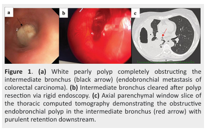
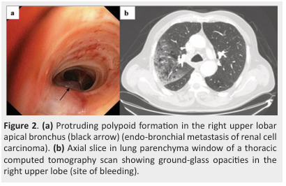
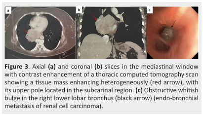
Cases: We presented three cases of EBMs, one secondary to colorectal cancer and two associated with renal tumours. EBM unveiled the extra-thoracic neoplasm in colorectal cancer and was incidentally discovered during renal cancer follow-up. Bronchoscopy revealed an obstructive endobronchial mass in two cases. Histological diagnosis was established via bronchial biopsies (collecting duct renal carcinoma), computed tomography-guided transparietal lung biopsy (clear cell renal carcinoma), and endobronchial mass resection through rigid bronchoscopy (colorectal adenocarcinoma).
Conclusion: In case of an endobronchial lesion, the diagnosis of EBM should be evoked especially when the medical history reports extra-thoracic neoplasms. This diagnostic hypothesis guides the histological diagnosis and leads to an appropriate treatment.

 求助内容:
求助内容: 应助结果提醒方式:


