{"title":"左髂总动脉瘤破裂至髂静脉并静脉异常1例报告。","authors":"Shunta Hayakawa, Jien Saito, Shinji Kamiya, Yoshiaki Sone, Yukihide Numata, Hideki Sasaki","doi":"10.3400/avd.cr.24-00027","DOIUrl":null,"url":null,"abstract":"<p><p>Ruptured iliac artery aneurysms are serious conditions with high mortality, occasionally perforating into the venous system. A 73-year-old male presented with left leg edema and a pulsatile left lower abdominal mass. Computed tomography revealed a ruptured left common iliac artery aneurysm with perforation into the left common iliac vein. Additionally, bilateral internal iliac veins were noted to form an anomalous common trunk draining into the left common iliac vein. Treatment involved internal iliac vein balloon occlusion under fluoroscopy followed by open surgery for artificial graft replacement and fistula repair. The patient was discharged on the 8th postoperative day.</p>","PeriodicalId":7995,"journal":{"name":"Annals of vascular diseases","volume":"17 4","pages":"413-416"},"PeriodicalIF":0.6000,"publicationDate":"2024-12-25","publicationTypes":"Journal Article","fieldsOfStudy":null,"isOpenAccess":false,"openAccessPdf":"https://www.ncbi.nlm.nih.gov/pmc/articles/PMC11669026/pdf/","citationCount":"0","resultStr":"{\"title\":\"Left Common Iliac Artery Aneurysm Rupture to an Iliac Vein with a Venous Anomaly: A Case Report.\",\"authors\":\"Shunta Hayakawa, Jien Saito, Shinji Kamiya, Yoshiaki Sone, Yukihide Numata, Hideki Sasaki\",\"doi\":\"10.3400/avd.cr.24-00027\",\"DOIUrl\":null,\"url\":null,\"abstract\":\"<p><p>Ruptured iliac artery aneurysms are serious conditions with high mortality, occasionally perforating into the venous system. A 73-year-old male presented with left leg edema and a pulsatile left lower abdominal mass. Computed tomography revealed a ruptured left common iliac artery aneurysm with perforation into the left common iliac vein. Additionally, bilateral internal iliac veins were noted to form an anomalous common trunk draining into the left common iliac vein. Treatment involved internal iliac vein balloon occlusion under fluoroscopy followed by open surgery for artificial graft replacement and fistula repair. The patient was discharged on the 8th postoperative day.</p>\",\"PeriodicalId\":7995,\"journal\":{\"name\":\"Annals of vascular diseases\",\"volume\":\"17 4\",\"pages\":\"413-416\"},\"PeriodicalIF\":0.6000,\"publicationDate\":\"2024-12-25\",\"publicationTypes\":\"Journal Article\",\"fieldsOfStudy\":null,\"isOpenAccess\":false,\"openAccessPdf\":\"https://www.ncbi.nlm.nih.gov/pmc/articles/PMC11669026/pdf/\",\"citationCount\":\"0\",\"resultStr\":null,\"platform\":\"Semanticscholar\",\"paperid\":null,\"PeriodicalName\":\"Annals of vascular diseases\",\"FirstCategoryId\":\"1085\",\"ListUrlMain\":\"https://doi.org/10.3400/avd.cr.24-00027\",\"RegionNum\":0,\"RegionCategory\":null,\"ArticlePicture\":[],\"TitleCN\":null,\"AbstractTextCN\":null,\"PMCID\":null,\"EPubDate\":\"2024/10/24 0:00:00\",\"PubModel\":\"Epub\",\"JCR\":\"Q4\",\"JCRName\":\"PERIPHERAL VASCULAR DISEASE\",\"Score\":null,\"Total\":0}","platform":"Semanticscholar","paperid":null,"PeriodicalName":"Annals of vascular diseases","FirstCategoryId":"1085","ListUrlMain":"https://doi.org/10.3400/avd.cr.24-00027","RegionNum":0,"RegionCategory":null,"ArticlePicture":[],"TitleCN":null,"AbstractTextCN":null,"PMCID":null,"EPubDate":"2024/10/24 0:00:00","PubModel":"Epub","JCR":"Q4","JCRName":"PERIPHERAL VASCULAR DISEASE","Score":null,"Total":0}
Left Common Iliac Artery Aneurysm Rupture to an Iliac Vein with a Venous Anomaly: A Case Report.
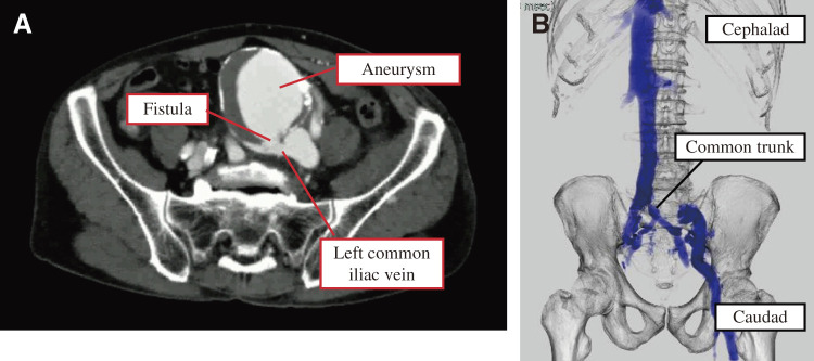
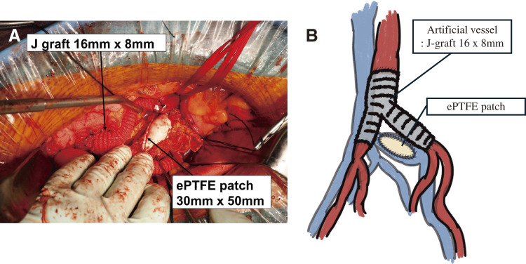
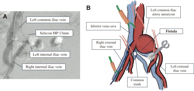
Ruptured iliac artery aneurysms are serious conditions with high mortality, occasionally perforating into the venous system. A 73-year-old male presented with left leg edema and a pulsatile left lower abdominal mass. Computed tomography revealed a ruptured left common iliac artery aneurysm with perforation into the left common iliac vein. Additionally, bilateral internal iliac veins were noted to form an anomalous common trunk draining into the left common iliac vein. Treatment involved internal iliac vein balloon occlusion under fluoroscopy followed by open surgery for artificial graft replacement and fistula repair. The patient was discharged on the 8th postoperative day.

 求助内容:
求助内容: 应助结果提醒方式:


