Sixuan Wu, Kefan Song, Jason Cobb, Alexander T Adams
{"title":"无泵微流体细胞浓度分析的智能手机在临床设置(SmartFlow):设计,开发和评估。","authors":"Sixuan Wu, Kefan Song, Jason Cobb, Alexander T Adams","doi":"10.2196/62770","DOIUrl":null,"url":null,"abstract":"<p><strong>Background: </strong>Cell concentration in body fluid is an important factor for clinical diagnosis. The traditional method involves clinicians manually counting cells under microscopes, which is labor-intensive. Automated cell concentration estimation can be achieved using flow cytometers; however, their high cost limits accessibility. Microfluidic systems, although cheaper than flow cytometers, still require high-speed cameras and syringe pumps to drive the flow and ensure video quality. In this paper, we present SmartFlow, a low-cost solution for cell concentration estimation using smartphone-based computer vision on 3D-printed, pump-free microfluidic platforms.</p><p><strong>Objective: </strong>The objective was to design and fabricate microfluidic chips, coupled with clinical utilities, for cell counting and concentration analysis. We answered the following research questions (RQs): RQ1, Can gravity drive the flow within the microfluidic chips, eliminating the need for external pumps? RQ2, How does the microfluidic chip design impact video quality for cell analysis? RQ3, Can smartphone-captured videos be used to estimate cell count and concentration in microfluidic chips?</p><p><strong>Methods: </strong>To answer the 3 RQs, 2 experiments were conducted. In the cell flow velocity experiment, diluted sheep blood flowed through the microfluidic chips with and without a bottleneck design to answer RQ1 and RQ2, respectively. In the cell concentration analysis experiment, sheep blood diluted into 13 concentrations flowed through the microfluidic chips while videos were recorded by smartphones for the concentration measurement.</p><p><strong>Results: </strong>In the cell flow velocity experiment, we designed and fabricated 2 versions of microfluidic chips. The ANOVA test (Straight: F<sub>6, 99</sub>=6144.45, P<.001; Bottleneck: F<sub>6, 99</sub>=3475.78, P<.001) showed the height difference had a significant impact on the cell velocity, which implied gravity could drive the flow. The video sharpness analysis demonstrated that video quality followed an exponential decay with increasing height differences (video quality=100e<sup>-k×Height</sup>) and a bottleneck design could effectively preserve video quality (Straight: R<sup>2</sup>=0.95, k=4.33; Bottleneck: R<sup>2</sup>=0.91, k=0.59). Samples from the 13 cell concentrations were used for cell counting and cell concentration estimation analysis. The accuracy of cell counting (n=35, 60-second samples, R<sup>2</sup>=0.96, mean absolute error=1.10, mean squared error=2.24, root mean squared error=1.50) and cell concentration regression (n=39, 150-second samples, R<sup>2</sup>=0.99, mean absolute error=0.24, mean squared error=0.11, root mean squared error=0.33 on a logarithmic scale, mean average percentage error=0.25) were evaluated using 5-fold cross-validation by comparing the algorithmic estimation to ground truth.</p><p><strong>Conclusions: </strong>In conclusion, we demonstrated the importance of the flow velocity in a microfluidic system, and we proposed SmartFlow, a low-cost system for computer vision-based cellular analysis. The proposed system could count the cells and estimate cell concentrations in the samples.</p>","PeriodicalId":87288,"journal":{"name":"JMIR biomedical engineering","volume":"9 ","pages":"e62770"},"PeriodicalIF":0.0000,"publicationDate":"2024-12-23","publicationTypes":"Journal Article","fieldsOfStudy":null,"isOpenAccess":false,"openAccessPdf":"https://www.ncbi.nlm.nih.gov/pmc/articles/PMC11704648/pdf/","citationCount":"0","resultStr":"{\"title\":\"Pump-Free Microfluidics for Cell Concentration Analysis on Smartphones in Clinical Settings (SmartFlow): Design, Development, and Evaluation.\",\"authors\":\"Sixuan Wu, Kefan Song, Jason Cobb, Alexander T Adams\",\"doi\":\"10.2196/62770\",\"DOIUrl\":null,\"url\":null,\"abstract\":\"<p><strong>Background: </strong>Cell concentration in body fluid is an important factor for clinical diagnosis. The traditional method involves clinicians manually counting cells under microscopes, which is labor-intensive. Automated cell concentration estimation can be achieved using flow cytometers; however, their high cost limits accessibility. Microfluidic systems, although cheaper than flow cytometers, still require high-speed cameras and syringe pumps to drive the flow and ensure video quality. In this paper, we present SmartFlow, a low-cost solution for cell concentration estimation using smartphone-based computer vision on 3D-printed, pump-free microfluidic platforms.</p><p><strong>Objective: </strong>The objective was to design and fabricate microfluidic chips, coupled with clinical utilities, for cell counting and concentration analysis. We answered the following research questions (RQs): RQ1, Can gravity drive the flow within the microfluidic chips, eliminating the need for external pumps? RQ2, How does the microfluidic chip design impact video quality for cell analysis? RQ3, Can smartphone-captured videos be used to estimate cell count and concentration in microfluidic chips?</p><p><strong>Methods: </strong>To answer the 3 RQs, 2 experiments were conducted. In the cell flow velocity experiment, diluted sheep blood flowed through the microfluidic chips with and without a bottleneck design to answer RQ1 and RQ2, respectively. In the cell concentration analysis experiment, sheep blood diluted into 13 concentrations flowed through the microfluidic chips while videos were recorded by smartphones for the concentration measurement.</p><p><strong>Results: </strong>In the cell flow velocity experiment, we designed and fabricated 2 versions of microfluidic chips. The ANOVA test (Straight: F<sub>6, 99</sub>=6144.45, P<.001; Bottleneck: F<sub>6, 99</sub>=3475.78, P<.001) showed the height difference had a significant impact on the cell velocity, which implied gravity could drive the flow. The video sharpness analysis demonstrated that video quality followed an exponential decay with increasing height differences (video quality=100e<sup>-k×Height</sup>) and a bottleneck design could effectively preserve video quality (Straight: R<sup>2</sup>=0.95, k=4.33; Bottleneck: R<sup>2</sup>=0.91, k=0.59). Samples from the 13 cell concentrations were used for cell counting and cell concentration estimation analysis. The accuracy of cell counting (n=35, 60-second samples, R<sup>2</sup>=0.96, mean absolute error=1.10, mean squared error=2.24, root mean squared error=1.50) and cell concentration regression (n=39, 150-second samples, R<sup>2</sup>=0.99, mean absolute error=0.24, mean squared error=0.11, root mean squared error=0.33 on a logarithmic scale, mean average percentage error=0.25) were evaluated using 5-fold cross-validation by comparing the algorithmic estimation to ground truth.</p><p><strong>Conclusions: </strong>In conclusion, we demonstrated the importance of the flow velocity in a microfluidic system, and we proposed SmartFlow, a low-cost system for computer vision-based cellular analysis. The proposed system could count the cells and estimate cell concentrations in the samples.</p>\",\"PeriodicalId\":87288,\"journal\":{\"name\":\"JMIR biomedical engineering\",\"volume\":\"9 \",\"pages\":\"e62770\"},\"PeriodicalIF\":0.0000,\"publicationDate\":\"2024-12-23\",\"publicationTypes\":\"Journal Article\",\"fieldsOfStudy\":null,\"isOpenAccess\":false,\"openAccessPdf\":\"https://www.ncbi.nlm.nih.gov/pmc/articles/PMC11704648/pdf/\",\"citationCount\":\"0\",\"resultStr\":null,\"platform\":\"Semanticscholar\",\"paperid\":null,\"PeriodicalName\":\"JMIR biomedical engineering\",\"FirstCategoryId\":\"1085\",\"ListUrlMain\":\"https://doi.org/10.2196/62770\",\"RegionNum\":0,\"RegionCategory\":null,\"ArticlePicture\":[],\"TitleCN\":null,\"AbstractTextCN\":null,\"PMCID\":null,\"EPubDate\":\"\",\"PubModel\":\"\",\"JCR\":\"\",\"JCRName\":\"\",\"Score\":null,\"Total\":0}","platform":"Semanticscholar","paperid":null,"PeriodicalName":"JMIR biomedical engineering","FirstCategoryId":"1085","ListUrlMain":"https://doi.org/10.2196/62770","RegionNum":0,"RegionCategory":null,"ArticlePicture":[],"TitleCN":null,"AbstractTextCN":null,"PMCID":null,"EPubDate":"","PubModel":"","JCR":"","JCRName":"","Score":null,"Total":0}
Pump-Free Microfluidics for Cell Concentration Analysis on Smartphones in Clinical Settings (SmartFlow): Design, Development, and Evaluation.
Background: Cell concentration in body fluid is an important factor for clinical diagnosis. The traditional method involves clinicians manually counting cells under microscopes, which is labor-intensive. Automated cell concentration estimation can be achieved using flow cytometers; however, their high cost limits accessibility. Microfluidic systems, although cheaper than flow cytometers, still require high-speed cameras and syringe pumps to drive the flow and ensure video quality. In this paper, we present SmartFlow, a low-cost solution for cell concentration estimation using smartphone-based computer vision on 3D-printed, pump-free microfluidic platforms.
Objective: The objective was to design and fabricate microfluidic chips, coupled with clinical utilities, for cell counting and concentration analysis. We answered the following research questions (RQs): RQ1, Can gravity drive the flow within the microfluidic chips, eliminating the need for external pumps? RQ2, How does the microfluidic chip design impact video quality for cell analysis? RQ3, Can smartphone-captured videos be used to estimate cell count and concentration in microfluidic chips?
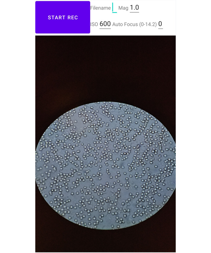
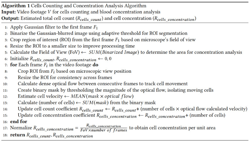
Methods: To answer the 3 RQs, 2 experiments were conducted. In the cell flow velocity experiment, diluted sheep blood flowed through the microfluidic chips with and without a bottleneck design to answer RQ1 and RQ2, respectively. In the cell concentration analysis experiment, sheep blood diluted into 13 concentrations flowed through the microfluidic chips while videos were recorded by smartphones for the concentration measurement.
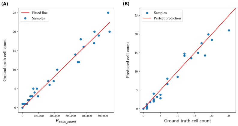
Results: In the cell flow velocity experiment, we designed and fabricated 2 versions of microfluidic chips. The ANOVA test (Straight: F6, 99=6144.45, P<.001; Bottleneck: F6, 99=3475.78, P<.001) showed the height difference had a significant impact on the cell velocity, which implied gravity could drive the flow. The video sharpness analysis demonstrated that video quality followed an exponential decay with increasing height differences (video quality=100e-k×Height) and a bottleneck design could effectively preserve video quality (Straight: R2=0.95, k=4.33; Bottleneck: R2=0.91, k=0.59). Samples from the 13 cell concentrations were used for cell counting and cell concentration estimation analysis. The accuracy of cell counting (n=35, 60-second samples, R2=0.96, mean absolute error=1.10, mean squared error=2.24, root mean squared error=1.50) and cell concentration regression (n=39, 150-second samples, R2=0.99, mean absolute error=0.24, mean squared error=0.11, root mean squared error=0.33 on a logarithmic scale, mean average percentage error=0.25) were evaluated using 5-fold cross-validation by comparing the algorithmic estimation to ground truth.
Conclusions: In conclusion, we demonstrated the importance of the flow velocity in a microfluidic system, and we proposed SmartFlow, a low-cost system for computer vision-based cellular analysis. The proposed system could count the cells and estimate cell concentrations in the samples.

 求助内容:
求助内容: 应助结果提醒方式:


