Atai Daniel, Matan Coronel, Segev Peer, Ben Grinshpan, Soner Duru, Jose L Peiro, James L Leach, Elena Abellán, Carolyn M Doerning, David Zarrouk, Francesco T Mangano
{"title":"一种新的微创神经外科颅固定装置,用于提高脑室内导管放置的准确性:实验动物研究。","authors":"Atai Daniel, Matan Coronel, Segev Peer, Ben Grinshpan, Soner Duru, Jose L Peiro, James L Leach, Elena Abellán, Carolyn M Doerning, David Zarrouk, Francesco T Mangano","doi":"10.1186/s13037-024-00420-0","DOIUrl":null,"url":null,"abstract":"<p><strong>Background: </strong>External ventricular drain (EVD) insertion is one of the most commonly performed neurosurgical procedures. Herein, we introduce a new concept of a cranial fixation device for insertion of EVDs, that reduces reliance on freehand placement and drilling techniques and provides a simple, minimally invasive approach that provides strong fixation to minimal thickness skulls.</p><p><strong>Methods: </strong>An experimental device for catheter insertion and fixation was designed and tested in both ex-vivo and in-vivo conditions to assess accurate cannulation of the ventricle and to test the strength of fixation to the skull. The ex-vivo experiments were conducted at Ben-Gurion University of the Negev (BGU) in Be'er Sheva, Israel. These experiments included functionality bench testing and pullout force measurements for the ball mechanism and catheter fixation. For the in-vivo experiments the fixation device was initially tested at the Cincinnati Children's Hospital Medical Center (CCHMC) in Cincinnati, Ohio on one day of life 1 (DOL 1) male control lamb. Additional experiments were conducted on 3 hydrocephalic DOL 0 lambs (1 male 2 female) at the Jesús Usón Minimally Invasive Surgery Centre (JUMISC) in Caceres, Spain. The hydrocephalic animal model used for this study was created with in utero intracisternal injection of BioGlue in fetal lambs. The catheter insertion trajectory was determined using MR imaging to assess the device's impact on the placement accuracy. The fixation device was evaluated on reaching the ventricle and enabling extraction of CSF for all 7 fixations placed. For 5 of the fixation devices, post-mortem pullout force was measured. The general functionality of the device was also evaluated.</p><p><strong>Results: </strong>In the experiments, 7/7 (100%) catheter trajectories successfully reached the ventricle without any apparent complications related to the device or the procedure. The cranial fixation device base demonstrated significant strength in withstanding an average pull-out force of 4.18kgf (STD[Formula: see text]0.72, N = 5) without detachment from the subject's skull for all 5 devices included in this test. Additionally, the EVD catheter pull test was conducted with the addition of a safety loop which did not allow movement of the EVD to a force of 3.6kgf. At this force the catheter tore but did not release from its fixation point.</p><p><strong>Conclusion: </strong>The newly designed experimental device demonstrates initial proof of concept from ex vivo and in vivo testing. It appears suitable for accurate ventricular catheter placement and cranial fixation.</p>","PeriodicalId":46782,"journal":{"name":"Patient Safety in Surgery","volume":"18 1","pages":"36"},"PeriodicalIF":2.1000,"publicationDate":"2024-12-18","publicationTypes":"Journal Article","fieldsOfStudy":null,"isOpenAccess":false,"openAccessPdf":"https://www.ncbi.nlm.nih.gov/pmc/articles/PMC11657085/pdf/","citationCount":"0","resultStr":"{\"title\":\"A novel minimally invasive neurosurgical cranial fixation device for improved accuracy of intraventricular catheter placement: an experimental animal study.\",\"authors\":\"Atai Daniel, Matan Coronel, Segev Peer, Ben Grinshpan, Soner Duru, Jose L Peiro, James L Leach, Elena Abellán, Carolyn M Doerning, David Zarrouk, Francesco T Mangano\",\"doi\":\"10.1186/s13037-024-00420-0\",\"DOIUrl\":null,\"url\":null,\"abstract\":\"<p><strong>Background: </strong>External ventricular drain (EVD) insertion is one of the most commonly performed neurosurgical procedures. Herein, we introduce a new concept of a cranial fixation device for insertion of EVDs, that reduces reliance on freehand placement and drilling techniques and provides a simple, minimally invasive approach that provides strong fixation to minimal thickness skulls.</p><p><strong>Methods: </strong>An experimental device for catheter insertion and fixation was designed and tested in both ex-vivo and in-vivo conditions to assess accurate cannulation of the ventricle and to test the strength of fixation to the skull. The ex-vivo experiments were conducted at Ben-Gurion University of the Negev (BGU) in Be'er Sheva, Israel. These experiments included functionality bench testing and pullout force measurements for the ball mechanism and catheter fixation. For the in-vivo experiments the fixation device was initially tested at the Cincinnati Children's Hospital Medical Center (CCHMC) in Cincinnati, Ohio on one day of life 1 (DOL 1) male control lamb. Additional experiments were conducted on 3 hydrocephalic DOL 0 lambs (1 male 2 female) at the Jesús Usón Minimally Invasive Surgery Centre (JUMISC) in Caceres, Spain. The hydrocephalic animal model used for this study was created with in utero intracisternal injection of BioGlue in fetal lambs. The catheter insertion trajectory was determined using MR imaging to assess the device's impact on the placement accuracy. The fixation device was evaluated on reaching the ventricle and enabling extraction of CSF for all 7 fixations placed. For 5 of the fixation devices, post-mortem pullout force was measured. The general functionality of the device was also evaluated.</p><p><strong>Results: </strong>In the experiments, 7/7 (100%) catheter trajectories successfully reached the ventricle without any apparent complications related to the device or the procedure. The cranial fixation device base demonstrated significant strength in withstanding an average pull-out force of 4.18kgf (STD[Formula: see text]0.72, N = 5) without detachment from the subject's skull for all 5 devices included in this test. Additionally, the EVD catheter pull test was conducted with the addition of a safety loop which did not allow movement of the EVD to a force of 3.6kgf. At this force the catheter tore but did not release from its fixation point.</p><p><strong>Conclusion: </strong>The newly designed experimental device demonstrates initial proof of concept from ex vivo and in vivo testing. It appears suitable for accurate ventricular catheter placement and cranial fixation.</p>\",\"PeriodicalId\":46782,\"journal\":{\"name\":\"Patient Safety in Surgery\",\"volume\":\"18 1\",\"pages\":\"36\"},\"PeriodicalIF\":2.1000,\"publicationDate\":\"2024-12-18\",\"publicationTypes\":\"Journal Article\",\"fieldsOfStudy\":null,\"isOpenAccess\":false,\"openAccessPdf\":\"https://www.ncbi.nlm.nih.gov/pmc/articles/PMC11657085/pdf/\",\"citationCount\":\"0\",\"resultStr\":null,\"platform\":\"Semanticscholar\",\"paperid\":null,\"PeriodicalName\":\"Patient Safety in Surgery\",\"FirstCategoryId\":\"1085\",\"ListUrlMain\":\"https://doi.org/10.1186/s13037-024-00420-0\",\"RegionNum\":0,\"RegionCategory\":null,\"ArticlePicture\":[],\"TitleCN\":null,\"AbstractTextCN\":null,\"PMCID\":null,\"EPubDate\":\"\",\"PubModel\":\"\",\"JCR\":\"Q1\",\"JCRName\":\"SURGERY\",\"Score\":null,\"Total\":0}","platform":"Semanticscholar","paperid":null,"PeriodicalName":"Patient Safety in Surgery","FirstCategoryId":"1085","ListUrlMain":"https://doi.org/10.1186/s13037-024-00420-0","RegionNum":0,"RegionCategory":null,"ArticlePicture":[],"TitleCN":null,"AbstractTextCN":null,"PMCID":null,"EPubDate":"","PubModel":"","JCR":"Q1","JCRName":"SURGERY","Score":null,"Total":0}
A novel minimally invasive neurosurgical cranial fixation device for improved accuracy of intraventricular catheter placement: an experimental animal study.
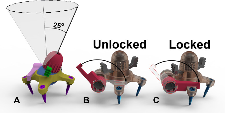
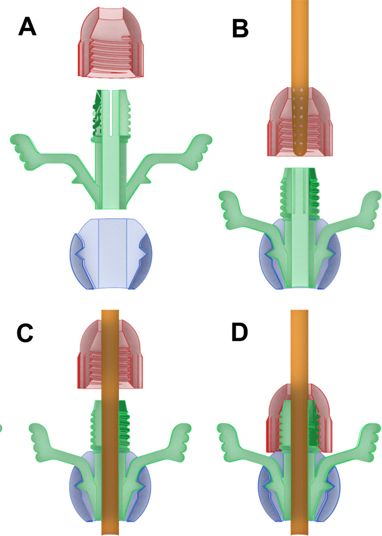
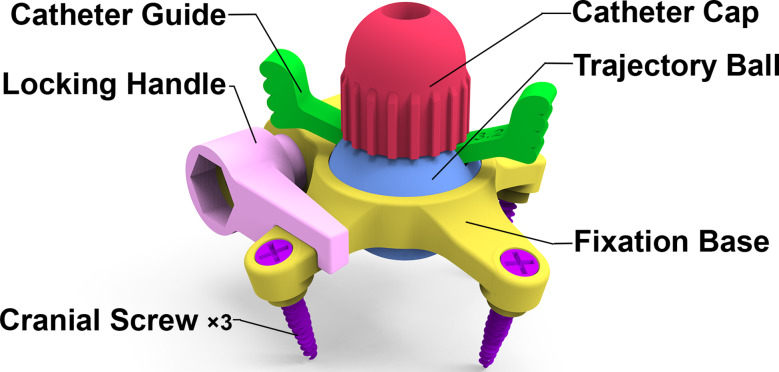
Background: External ventricular drain (EVD) insertion is one of the most commonly performed neurosurgical procedures. Herein, we introduce a new concept of a cranial fixation device for insertion of EVDs, that reduces reliance on freehand placement and drilling techniques and provides a simple, minimally invasive approach that provides strong fixation to minimal thickness skulls.
Methods: An experimental device for catheter insertion and fixation was designed and tested in both ex-vivo and in-vivo conditions to assess accurate cannulation of the ventricle and to test the strength of fixation to the skull. The ex-vivo experiments were conducted at Ben-Gurion University of the Negev (BGU) in Be'er Sheva, Israel. These experiments included functionality bench testing and pullout force measurements for the ball mechanism and catheter fixation. For the in-vivo experiments the fixation device was initially tested at the Cincinnati Children's Hospital Medical Center (CCHMC) in Cincinnati, Ohio on one day of life 1 (DOL 1) male control lamb. Additional experiments were conducted on 3 hydrocephalic DOL 0 lambs (1 male 2 female) at the Jesús Usón Minimally Invasive Surgery Centre (JUMISC) in Caceres, Spain. The hydrocephalic animal model used for this study was created with in utero intracisternal injection of BioGlue in fetal lambs. The catheter insertion trajectory was determined using MR imaging to assess the device's impact on the placement accuracy. The fixation device was evaluated on reaching the ventricle and enabling extraction of CSF for all 7 fixations placed. For 5 of the fixation devices, post-mortem pullout force was measured. The general functionality of the device was also evaluated.
Results: In the experiments, 7/7 (100%) catheter trajectories successfully reached the ventricle without any apparent complications related to the device or the procedure. The cranial fixation device base demonstrated significant strength in withstanding an average pull-out force of 4.18kgf (STD[Formula: see text]0.72, N = 5) without detachment from the subject's skull for all 5 devices included in this test. Additionally, the EVD catheter pull test was conducted with the addition of a safety loop which did not allow movement of the EVD to a force of 3.6kgf. At this force the catheter tore but did not release from its fixation point.
Conclusion: The newly designed experimental device demonstrates initial proof of concept from ex vivo and in vivo testing. It appears suitable for accurate ventricular catheter placement and cranial fixation.

 求助内容:
求助内容: 应助结果提醒方式:


