{"title":"右心室导联穿孔致心包气肿的发生机制及治疗。","authors":"Tomo Komaki, Yuuki Ueno, Noriyuki Mohri, Akihito Ideishi, Kohei Tashiro, Shin-Ichiro Miura, Masahiro Ogawa","doi":"10.14740/cr1738","DOIUrl":null,"url":null,"abstract":"<p><p>An 83-year-old man underwent dual-chamber pacemaker placement for complete atrioventricular block at another hospital. The active-fixation ventricular lead was positioned on the free wall of the anterior right ventricle. Ventricular pacing failure occurred on the day after pacemaker implantation, and fluoroscopy revealed right ventricular (RV) lead perforation. The patient was transferred to our hospital, and chest computed tomography revealed a severe pneumothorax and moderate pneumopericardium. These symptoms were relieved after chest tube drainage, and the patient's hemodynamics stabilized. The RV lead was percutaneously removed using simple traction under fluoroscopic guidance with cardiac surgical backup and was uneventfully refixed to the RV septum. Although there have been several reports of pneumopericardium caused by atrial lead perforation, there are very few cases related to RV lead. Pneumopericardium complicated by pneumothorax due to RV lead perforation can be relieved using chest tube drainage without the need for pericardiocentesis.</p>","PeriodicalId":9424,"journal":{"name":"Cardiology Research","volume":"15 6","pages":"472-476"},"PeriodicalIF":1.4000,"publicationDate":"2024-12-01","publicationTypes":"Journal Article","fieldsOfStudy":null,"isOpenAccess":false,"openAccessPdf":"https://www.ncbi.nlm.nih.gov/pmc/articles/PMC11650576/pdf/","citationCount":"0","resultStr":"{\"title\":\"The Mechanism and Management of Pneumopericardium Caused by Right Ventricular Lead Perforation.\",\"authors\":\"Tomo Komaki, Yuuki Ueno, Noriyuki Mohri, Akihito Ideishi, Kohei Tashiro, Shin-Ichiro Miura, Masahiro Ogawa\",\"doi\":\"10.14740/cr1738\",\"DOIUrl\":null,\"url\":null,\"abstract\":\"<p><p>An 83-year-old man underwent dual-chamber pacemaker placement for complete atrioventricular block at another hospital. The active-fixation ventricular lead was positioned on the free wall of the anterior right ventricle. Ventricular pacing failure occurred on the day after pacemaker implantation, and fluoroscopy revealed right ventricular (RV) lead perforation. The patient was transferred to our hospital, and chest computed tomography revealed a severe pneumothorax and moderate pneumopericardium. These symptoms were relieved after chest tube drainage, and the patient's hemodynamics stabilized. The RV lead was percutaneously removed using simple traction under fluoroscopic guidance with cardiac surgical backup and was uneventfully refixed to the RV septum. Although there have been several reports of pneumopericardium caused by atrial lead perforation, there are very few cases related to RV lead. Pneumopericardium complicated by pneumothorax due to RV lead perforation can be relieved using chest tube drainage without the need for pericardiocentesis.</p>\",\"PeriodicalId\":9424,\"journal\":{\"name\":\"Cardiology Research\",\"volume\":\"15 6\",\"pages\":\"472-476\"},\"PeriodicalIF\":1.4000,\"publicationDate\":\"2024-12-01\",\"publicationTypes\":\"Journal Article\",\"fieldsOfStudy\":null,\"isOpenAccess\":false,\"openAccessPdf\":\"https://www.ncbi.nlm.nih.gov/pmc/articles/PMC11650576/pdf/\",\"citationCount\":\"0\",\"resultStr\":null,\"platform\":\"Semanticscholar\",\"paperid\":null,\"PeriodicalName\":\"Cardiology Research\",\"FirstCategoryId\":\"1085\",\"ListUrlMain\":\"https://doi.org/10.14740/cr1738\",\"RegionNum\":0,\"RegionCategory\":null,\"ArticlePicture\":[],\"TitleCN\":null,\"AbstractTextCN\":null,\"PMCID\":null,\"EPubDate\":\"2024/12/3 0:00:00\",\"PubModel\":\"Epub\",\"JCR\":\"Q3\",\"JCRName\":\"CARDIAC & CARDIOVASCULAR SYSTEMS\",\"Score\":null,\"Total\":0}","platform":"Semanticscholar","paperid":null,"PeriodicalName":"Cardiology Research","FirstCategoryId":"1085","ListUrlMain":"https://doi.org/10.14740/cr1738","RegionNum":0,"RegionCategory":null,"ArticlePicture":[],"TitleCN":null,"AbstractTextCN":null,"PMCID":null,"EPubDate":"2024/12/3 0:00:00","PubModel":"Epub","JCR":"Q3","JCRName":"CARDIAC & CARDIOVASCULAR SYSTEMS","Score":null,"Total":0}
The Mechanism and Management of Pneumopericardium Caused by Right Ventricular Lead Perforation.
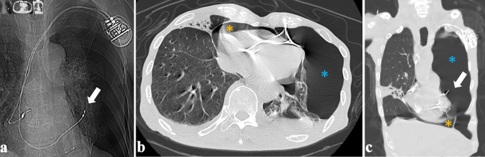
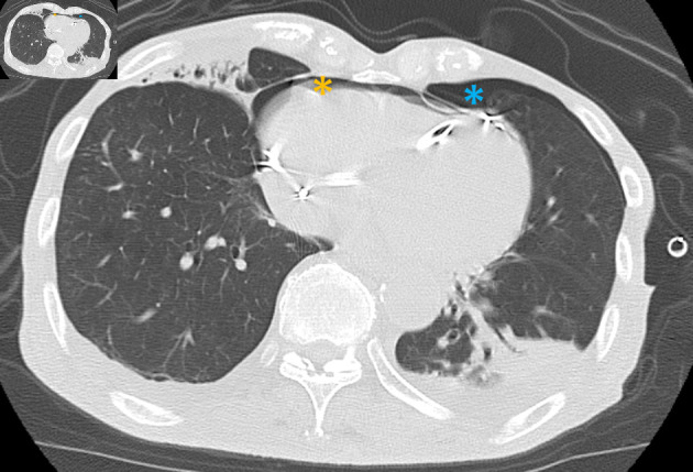
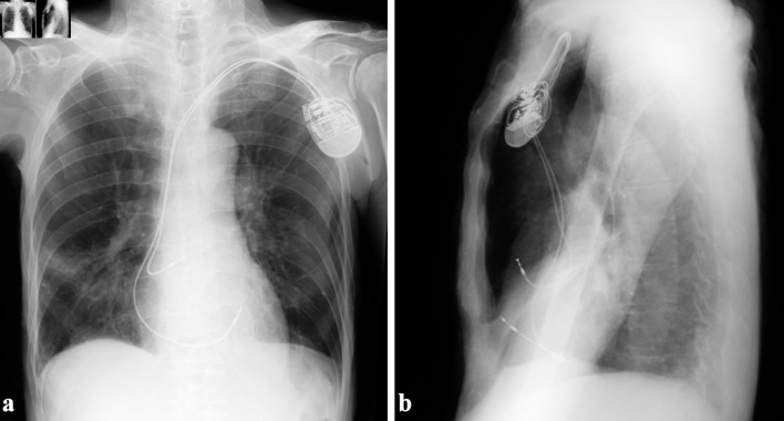
An 83-year-old man underwent dual-chamber pacemaker placement for complete atrioventricular block at another hospital. The active-fixation ventricular lead was positioned on the free wall of the anterior right ventricle. Ventricular pacing failure occurred on the day after pacemaker implantation, and fluoroscopy revealed right ventricular (RV) lead perforation. The patient was transferred to our hospital, and chest computed tomography revealed a severe pneumothorax and moderate pneumopericardium. These symptoms were relieved after chest tube drainage, and the patient's hemodynamics stabilized. The RV lead was percutaneously removed using simple traction under fluoroscopic guidance with cardiac surgical backup and was uneventfully refixed to the RV septum. Although there have been several reports of pneumopericardium caused by atrial lead perforation, there are very few cases related to RV lead. Pneumopericardium complicated by pneumothorax due to RV lead perforation can be relieved using chest tube drainage without the need for pericardiocentesis.
期刊介绍:
Cardiology Research is an open access, peer-reviewed, international journal. All submissions relating to basic research and clinical practice of cardiology and cardiovascular medicine are in this journal''s scope. This journal focuses on publishing original research and observations in all cardiovascular medicine aspects. Manuscript types include original article, review, case report, short communication, book review, letter to the editor.

 求助内容:
求助内容: 应助结果提醒方式:


