经典霍奇金淋巴瘤肿瘤微环境中的 T 细胞多样性和血源性 T 细胞排斥作用

IF 12.8 1区 医学 Q1 HEMATOLOGY
Nicole Seifert, Sarah Reinke, Johanna Grund, Berit Müller-Meinhard, Julia Richter, Thorsten Heilmann, Hans Schlößer, Michaela Kotrova, Monika Brüggemann, Peter Borchmann, Paul J. Bröckelmann, Michael Altenbuchinger, Wolfram Klapper
{"title":"经典霍奇金淋巴瘤肿瘤微环境中的 T 细胞多样性和血源性 T 细胞排斥作用","authors":"Nicole Seifert, Sarah Reinke, Johanna Grund, Berit Müller-Meinhard, Julia Richter, Thorsten Heilmann, Hans Schlößer, Michaela Kotrova, Monika Brüggemann, Peter Borchmann, Paul J. Bröckelmann, Michael Altenbuchinger, Wolfram Klapper","doi":"10.1038/s41375-024-02490-6","DOIUrl":null,"url":null,"abstract":"<p>The Tumor Microenvironment (TME) in classical Hodgkin Lymphoma (HL) contains abundant immune cells and only few neoplastic Hodgkin and Reed-Sternberg cells (HRSC). We analyzed the T-cell receptor (TCR) repertoire to detect T-cell expansion in the TME and blood. In contrast to solid cancer tissue, T-cells in the TME of HL are highly polyclonal at first diagnosis and show only minor clonal expansion during anti-PD1 immune checkpoint blockade (ICB). At relapse and during ICB, pre-amplified T-cell populations increase in the TME of solid cancers but to a much lesser extent in HL. In contrast, T-cell populations in the peripheral blood of HL patients display higher clonality than healthy controls reaching clonality levels comparable to solid cancer. However, pre-amplified blood T-cells in HL patients show only minor additional clonal expansion during ICB. Moreover, blood-derived T-cells do not repopulate the TME of HL to the same extent as observed in solid cancers. Thus, the T-cell repertoire in the TME of HL appears unique by a relatively low clonal T-cell content and the exclusion of clonally expanded T-cells from the peripheral blood. Exclusion of clonally expanded tumor-specific T-cells from the TME may present a novel mechanism of immune evasion in HL.</p>","PeriodicalId":18109,"journal":{"name":"Leukemia","volume":"45 1","pages":""},"PeriodicalIF":12.8000,"publicationDate":"2024-12-17","publicationTypes":"Journal Article","fieldsOfStudy":null,"isOpenAccess":false,"openAccessPdf":"","citationCount":"0","resultStr":"{\"title\":\"T-cell diversity and exclusion of blood-derived T-cells in the tumor microenvironment of classical Hodgkin Lymphoma\",\"authors\":\"Nicole Seifert, Sarah Reinke, Johanna Grund, Berit Müller-Meinhard, Julia Richter, Thorsten Heilmann, Hans Schlößer, Michaela Kotrova, Monika Brüggemann, Peter Borchmann, Paul J. Bröckelmann, Michael Altenbuchinger, Wolfram Klapper\",\"doi\":\"10.1038/s41375-024-02490-6\",\"DOIUrl\":null,\"url\":null,\"abstract\":\"<p>The Tumor Microenvironment (TME) in classical Hodgkin Lymphoma (HL) contains abundant immune cells and only few neoplastic Hodgkin and Reed-Sternberg cells (HRSC). We analyzed the T-cell receptor (TCR) repertoire to detect T-cell expansion in the TME and blood. In contrast to solid cancer tissue, T-cells in the TME of HL are highly polyclonal at first diagnosis and show only minor clonal expansion during anti-PD1 immune checkpoint blockade (ICB). At relapse and during ICB, pre-amplified T-cell populations increase in the TME of solid cancers but to a much lesser extent in HL. In contrast, T-cell populations in the peripheral blood of HL patients display higher clonality than healthy controls reaching clonality levels comparable to solid cancer. However, pre-amplified blood T-cells in HL patients show only minor additional clonal expansion during ICB. Moreover, blood-derived T-cells do not repopulate the TME of HL to the same extent as observed in solid cancers. Thus, the T-cell repertoire in the TME of HL appears unique by a relatively low clonal T-cell content and the exclusion of clonally expanded T-cells from the peripheral blood. Exclusion of clonally expanded tumor-specific T-cells from the TME may present a novel mechanism of immune evasion in HL.</p>\",\"PeriodicalId\":18109,\"journal\":{\"name\":\"Leukemia\",\"volume\":\"45 1\",\"pages\":\"\"},\"PeriodicalIF\":12.8000,\"publicationDate\":\"2024-12-17\",\"publicationTypes\":\"Journal Article\",\"fieldsOfStudy\":null,\"isOpenAccess\":false,\"openAccessPdf\":\"\",\"citationCount\":\"0\",\"resultStr\":null,\"platform\":\"Semanticscholar\",\"paperid\":null,\"PeriodicalName\":\"Leukemia\",\"FirstCategoryId\":\"3\",\"ListUrlMain\":\"https://doi.org/10.1038/s41375-024-02490-6\",\"RegionNum\":1,\"RegionCategory\":\"医学\",\"ArticlePicture\":[],\"TitleCN\":null,\"AbstractTextCN\":null,\"PMCID\":null,\"EPubDate\":\"\",\"PubModel\":\"\",\"JCR\":\"Q1\",\"JCRName\":\"HEMATOLOGY\",\"Score\":null,\"Total\":0}","platform":"Semanticscholar","paperid":null,"PeriodicalName":"Leukemia","FirstCategoryId":"3","ListUrlMain":"https://doi.org/10.1038/s41375-024-02490-6","RegionNum":1,"RegionCategory":"医学","ArticlePicture":[],"TitleCN":null,"AbstractTextCN":null,"PMCID":null,"EPubDate":"","PubModel":"","JCR":"Q1","JCRName":"HEMATOLOGY","Score":null,"Total":0}
引用次数: 0

摘要

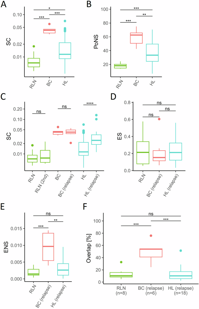
经典霍奇金淋巴瘤(HL)的肿瘤微环境(TME)中含有大量免疫细胞,只有少数肿瘤性霍奇金细胞和里德-斯特恩伯格细胞(HRSC)。我们分析了T细胞受体(TCR)谱系,以检测TME和血液中T细胞的扩增情况。与实体瘤组织不同,HL的TME中的T细胞在初诊时高度多克隆,在抗PD1免疫检查点阻断(ICB)期间仅表现出轻微的克隆扩增。在复发和 ICB 期间,实体瘤 TME 中的预扩增 T 细胞群会增加,但在 HL 中增加的程度要小得多。相比之下,HL 患者外周血中的 T 细胞群的克隆度高于健康对照组,达到了与实体癌相当的克隆度水平。然而,HL 患者的预扩增血液 T 细胞在 ICB 期间仅表现出轻微的额外克隆扩增。此外,血液中的 T 细胞在 HL 的 TME 中的重新填充程度与实体瘤中观察到的不同。因此,HL 的 TME 中的 T 细胞谱似乎是独特的,其克隆 T 细胞含量相对较低,外周血中排除了克隆扩增的 T 细胞。将克隆扩增的肿瘤特异性T细胞排除在TME之外可能是HL免疫逃避的一种新机制。
本文章由计算机程序翻译,如有差异,请以英文原文为准。

T-cell diversity and exclusion of blood-derived T-cells in the tumor microenvironment of classical Hodgkin Lymphoma

T-cell diversity and exclusion of blood-derived T-cells in the tumor microenvironment of classical Hodgkin Lymphoma

The Tumor Microenvironment (TME) in classical Hodgkin Lymphoma (HL) contains abundant immune cells and only few neoplastic Hodgkin and Reed-Sternberg cells (HRSC). We analyzed the T-cell receptor (TCR) repertoire to detect T-cell expansion in the TME and blood. In contrast to solid cancer tissue, T-cells in the TME of HL are highly polyclonal at first diagnosis and show only minor clonal expansion during anti-PD1 immune checkpoint blockade (ICB). At relapse and during ICB, pre-amplified T-cell populations increase in the TME of solid cancers but to a much lesser extent in HL. In contrast, T-cell populations in the peripheral blood of HL patients display higher clonality than healthy controls reaching clonality levels comparable to solid cancer. However, pre-amplified blood T-cells in HL patients show only minor additional clonal expansion during ICB. Moreover, blood-derived T-cells do not repopulate the TME of HL to the same extent as observed in solid cancers. Thus, the T-cell repertoire in the TME of HL appears unique by a relatively low clonal T-cell content and the exclusion of clonally expanded T-cells from the peripheral blood. Exclusion of clonally expanded tumor-specific T-cells from the TME may present a novel mechanism of immune evasion in HL.

求助全文
通过发布文献求助,成功后即可免费获取论文全文。 去求助
来源期刊
Leukemia
Leukemia 医学-血液学
CiteScore
18.10
自引率
3.50%
发文量
270
审稿时长
3-6 weeks
期刊介绍: Title: Leukemia Journal Overview: Publishes high-quality, peer-reviewed research Covers all aspects of research and treatment of leukemia and allied diseases Includes studies of normal hemopoiesis due to comparative relevance Topics of Interest: Oncogenes Growth factors Stem cells Leukemia genomics Cell cycle Signal transduction Molecular targets for therapy And more Content Types: Original research articles Reviews Letters Correspondence Comments elaborating on significant advances and covering topical issues
×
引用
GB/T 7714-2015
复制
MLA
复制
APA
复制
导出至
BibTeX EndNote RefMan NoteFirst NoteExpress
×
提示
您的信息不完整,为了账户安全,请先补充。
现在去补充
×
提示
您因"违规操作"
具体请查看互助需知
我知道了
×
提示
确定
请完成安全验证×
copy
已复制链接
快去分享给好友吧!
我知道了
右上角分享
点击右上角分享
0
联系我们:info@booksci.cn Book学术提供免费学术资源搜索服务,方便国内外学者检索中英文文献。致力于提供最便捷和优质的服务体验。 Copyright © 2023 布克学术 All rights reserved.
京ICP备2023020795号-1
ghs 京公网安备 11010802042870号
Book学术文献互助
Book学术文献互助群
群 号:481959085
Book学术官方微信