修正“用x射线光电子能谱定量测定氧化锡/金核-壳纳米粒子的壳层厚度”
IF 3.2
3区 化学
Q2 CHEMISTRY, PHYSICAL
引用次数: 0
摘要
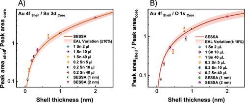
图 5.使用 SESSA v2.0 和实验 SnO@Au CSNP 样品获得的模拟 XPS 峰强比与纳米粒子外壳厚度的函数关系对比。(A) Au 4f/Sn 3d;(B) Au 4f/O 1s。图 S1.SnO@Au 核壳纳米粒子 (A) O 1s 和 (B) 勘测光谱区域的高分辨率 XPS 扫描。(A) 试验性 SnO@Au 和 SESSA 模拟核级光谱显示了氧化硅和氧化锡的模拟峰值。(B) 具有固定 1 纳米金壳的五种不同内核直径。SnO@Au 核壳 NPs 的 S1 SESSA V2.0 光谱:图 S1(a) SnO@Au 核壳 NPs 的 S1 SESSA V2.0 光谱:表 S1:模拟参数 本文引用了 1 篇其他出版物。本文尚未被其他出版物引用。本文章由计算机程序翻译,如有差异,请以英文原文为准。
Correction to “Quantification of the Shell Thickness of Tin Oxide/Gold Core–Shell Nanoparticles by X-ray Photoelectron Spectroscopy”
Figure 5. Comparison of simulated XPS peak intensity ratios as a function of the nanoparticle shell thickness obtained using SESSA v2.0 and experimental SnO@Au CSNP samples. (A) Au 4f/Sn 3d; (B) Au 4f/O 1s. Figure S1. High-resolution XPS scans in the region of (A) O 1s and (B) survey spectra of SnO@Au core–shell nanoparticles. (A) Experimental SnO@Au and SESSA simulated core level spectra showing simulated peaks from both silicon oxide and tin oxide contributions. (B) Five different core diameters with a fixed 1 nm gold shell. S1 SESSA V2.0 spectra of SnO@Au core–shell NPs: Figure S1(a) S1 SESSA V2.0 spectra of SnO@Au core–shell NPs: Table S1: Simulation parameters This article references 1 other publications. This article has not yet been cited by other publications.
求助全文
通过发布文献求助,成功后即可免费获取论文全文。
去求助
来源期刊
The Journal of Physical Chemistry C
化学-材料科学:综合
CiteScore
6.50
自引率
8.10%
发文量
2047
审稿时长
1.8 months
期刊介绍:
The Journal of Physical Chemistry A/B/C is devoted to reporting new and original experimental and theoretical basic research of interest to physical chemists, biophysical chemists, and chemical physicists.
 求助内容:
求助内容: 应助结果提醒方式:


