{"title":"开发使用3d打印模型的全内窥镜手术新手术培训。","authors":"Takahiro Ogawa, Masatoshi Morimoto, Shutaro Fujimoto, Masaru Tominaga, Yasuyuki Omichi, Kosuke Sugiura, Fumitake Tezuka, Kazuta Yamashita, Koichi Sairyo","doi":"10.22603/ssrr.2023-0285","DOIUrl":null,"url":null,"abstract":"<p><strong>Introduction: </strong>Full endoscopic spine surgery continues to spread worldwide but has a long learning curve. Conventional endoscopy training uses live pigs or human cadavers, which has disadvantages such as high costs and limited availability. Therefore, this study aimed to develop and evaluate three-dimensional (3D)-printed models for endoscopy training.</p><p><strong>Methods: </strong>Models for 3D printing were generated using raw imaging data from 1.0-mm slices of computed tomography scans, and each part was printed using a different colored material. The combined model was used for training as part of the full endoscopy training kit.</p><p><strong>Results: </strong>This approach offers several advantages. First, it enables the creation of accurate disease models, such as lumbar disc herniation and other abnormalities, which are useful for both surgical training and preoperative simulations. Second, it is useful for learning surgical orientation. During surgical training, the surgical field can be viewed directly through an endoscope or with the naked eye. By using various colors, it becomes easier to recognize the orientation. Third, the amount of drilling resection can be easily confirmed, facilitating feedback. Finally, training for various surgical techniques is possible, including endoscopic holding techniques and using the endoscope's outer sheath to retract nerves. However, this approach also has some disadvantages, such as the lack of bleeding, inability to reproduce tissue hardness, and difficulty in faithfully recreating soft tissue, such as connective tissue, blood vessels, and fat. Therefore, it is difficult to reproduce the hardness of the calcified disc or disc herniation with apophyseal ring fracture. Moreover, 3D-printed models are not suitable for surgical training using the interlaminal approach because it is difficult to perform separation between the ligamentum flavum and dural matter or between the dural matter and intervertebral disc.</p><p><strong>Conclusions: </strong>3D-printed models are a useful complement to live pigs and human cadavers in surgical training and can reduce the time required to acquire endoscopic skills.</p>","PeriodicalId":22253,"journal":{"name":"Spine Surgery and Related Research","volume":"8 6","pages":"591-599"},"PeriodicalIF":1.2000,"publicationDate":"2024-04-03","publicationTypes":"Journal Article","fieldsOfStudy":null,"isOpenAccess":false,"openAccessPdf":"https://www.ncbi.nlm.nih.gov/pmc/articles/PMC11625712/pdf/","citationCount":"0","resultStr":"{\"title\":\"Development of New Surgical Training for Full Endoscopic Surgery Using 3D-Printed Models.\",\"authors\":\"Takahiro Ogawa, Masatoshi Morimoto, Shutaro Fujimoto, Masaru Tominaga, Yasuyuki Omichi, Kosuke Sugiura, Fumitake Tezuka, Kazuta Yamashita, Koichi Sairyo\",\"doi\":\"10.22603/ssrr.2023-0285\",\"DOIUrl\":null,\"url\":null,\"abstract\":\"<p><strong>Introduction: </strong>Full endoscopic spine surgery continues to spread worldwide but has a long learning curve. Conventional endoscopy training uses live pigs or human cadavers, which has disadvantages such as high costs and limited availability. Therefore, this study aimed to develop and evaluate three-dimensional (3D)-printed models for endoscopy training.</p><p><strong>Methods: </strong>Models for 3D printing were generated using raw imaging data from 1.0-mm slices of computed tomography scans, and each part was printed using a different colored material. The combined model was used for training as part of the full endoscopy training kit.</p><p><strong>Results: </strong>This approach offers several advantages. First, it enables the creation of accurate disease models, such as lumbar disc herniation and other abnormalities, which are useful for both surgical training and preoperative simulations. Second, it is useful for learning surgical orientation. During surgical training, the surgical field can be viewed directly through an endoscope or with the naked eye. By using various colors, it becomes easier to recognize the orientation. Third, the amount of drilling resection can be easily confirmed, facilitating feedback. Finally, training for various surgical techniques is possible, including endoscopic holding techniques and using the endoscope's outer sheath to retract nerves. However, this approach also has some disadvantages, such as the lack of bleeding, inability to reproduce tissue hardness, and difficulty in faithfully recreating soft tissue, such as connective tissue, blood vessels, and fat. Therefore, it is difficult to reproduce the hardness of the calcified disc or disc herniation with apophyseal ring fracture. Moreover, 3D-printed models are not suitable for surgical training using the interlaminal approach because it is difficult to perform separation between the ligamentum flavum and dural matter or between the dural matter and intervertebral disc.</p><p><strong>Conclusions: </strong>3D-printed models are a useful complement to live pigs and human cadavers in surgical training and can reduce the time required to acquire endoscopic skills.</p>\",\"PeriodicalId\":22253,\"journal\":{\"name\":\"Spine Surgery and Related Research\",\"volume\":\"8 6\",\"pages\":\"591-599\"},\"PeriodicalIF\":1.2000,\"publicationDate\":\"2024-04-03\",\"publicationTypes\":\"Journal Article\",\"fieldsOfStudy\":null,\"isOpenAccess\":false,\"openAccessPdf\":\"https://www.ncbi.nlm.nih.gov/pmc/articles/PMC11625712/pdf/\",\"citationCount\":\"0\",\"resultStr\":null,\"platform\":\"Semanticscholar\",\"paperid\":null,\"PeriodicalName\":\"Spine Surgery and Related Research\",\"FirstCategoryId\":\"1085\",\"ListUrlMain\":\"https://doi.org/10.22603/ssrr.2023-0285\",\"RegionNum\":0,\"RegionCategory\":null,\"ArticlePicture\":[],\"TitleCN\":null,\"AbstractTextCN\":null,\"PMCID\":null,\"EPubDate\":\"2024/11/27 0:00:00\",\"PubModel\":\"eCollection\",\"JCR\":\"Q3\",\"JCRName\":\"SURGERY\",\"Score\":null,\"Total\":0}","platform":"Semanticscholar","paperid":null,"PeriodicalName":"Spine Surgery and Related Research","FirstCategoryId":"1085","ListUrlMain":"https://doi.org/10.22603/ssrr.2023-0285","RegionNum":0,"RegionCategory":null,"ArticlePicture":[],"TitleCN":null,"AbstractTextCN":null,"PMCID":null,"EPubDate":"2024/11/27 0:00:00","PubModel":"eCollection","JCR":"Q3","JCRName":"SURGERY","Score":null,"Total":0}
Development of New Surgical Training for Full Endoscopic Surgery Using 3D-Printed Models.
Introduction: Full endoscopic spine surgery continues to spread worldwide but has a long learning curve. Conventional endoscopy training uses live pigs or human cadavers, which has disadvantages such as high costs and limited availability. Therefore, this study aimed to develop and evaluate three-dimensional (3D)-printed models for endoscopy training.
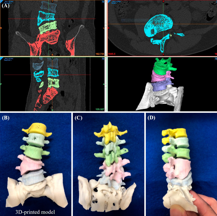
Methods: Models for 3D printing were generated using raw imaging data from 1.0-mm slices of computed tomography scans, and each part was printed using a different colored material. The combined model was used for training as part of the full endoscopy training kit.
Results: This approach offers several advantages. First, it enables the creation of accurate disease models, such as lumbar disc herniation and other abnormalities, which are useful for both surgical training and preoperative simulations. Second, it is useful for learning surgical orientation. During surgical training, the surgical field can be viewed directly through an endoscope or with the naked eye. By using various colors, it becomes easier to recognize the orientation. Third, the amount of drilling resection can be easily confirmed, facilitating feedback. Finally, training for various surgical techniques is possible, including endoscopic holding techniques and using the endoscope's outer sheath to retract nerves. However, this approach also has some disadvantages, such as the lack of bleeding, inability to reproduce tissue hardness, and difficulty in faithfully recreating soft tissue, such as connective tissue, blood vessels, and fat. Therefore, it is difficult to reproduce the hardness of the calcified disc or disc herniation with apophyseal ring fracture. Moreover, 3D-printed models are not suitable for surgical training using the interlaminal approach because it is difficult to perform separation between the ligamentum flavum and dural matter or between the dural matter and intervertebral disc.
Conclusions: 3D-printed models are a useful complement to live pigs and human cadavers in surgical training and can reduce the time required to acquire endoscopic skills.

 求助内容:
求助内容: 应助结果提醒方式:


