Anand Naranbhai, Amir Afrogheh, Suzanne O'Hagan, Johan Grobbelaar, Leon Janse van Rensburg
{"title":"南非一家医院hpv阳性与hpv阴性OPSCC的放射学特征","authors":"Anand Naranbhai, Amir Afrogheh, Suzanne O'Hagan, Johan Grobbelaar, Leon Janse van Rensburg","doi":"10.4102/sajr.v28i1.2976","DOIUrl":null,"url":null,"abstract":"<p><strong>Background: </strong>Studies have found that, at presentation, human papillomavirus (HPV)-positive oropharyngeal squamous cell carcinoma (OPSCC) has a less advanced primary tumour, more advanced lymph node spread and commonly has cystic metastatic lymph nodes in comparison to HPV-negative OPSCC.</p><p><strong>Objectives: </strong>To compare the radiological features of HPV-positive and HPV-negative OPSCC in South African patients.</p><p><strong>Method: </strong>A retrospective cross-sectional study was conducted at a large South African hospital. Eligibility required a histologically proven OPSCC between 2007 and 2023; a p16 antigen test and, if positive, a confirmatory HPV DNA PCR test and a baseline pre-treatment contrast enhanced neck CT scan. All eligible HPV-positive OPSCC patients and a random sample of eligible HPV-negative OPSCC patients were enrolled.</p><p><strong>Results: </strong>Twenty-one HPV-positive and 55 HPV-negative OPSCC patients were recruited. There was no statistically significant difference in the tumour epicentre location, local advancement (≥ T3 in 67% and 71%, respectively, <i>p</i> = 0.54), mean primary tumour size (41 mm vs. 39 mm, <i>p</i> = 0.73), lymph node spread (bilateral or more in 67% vs. 82%, <i>p</i> = 0.22) or morphologically cystic lymph nodes (10% and 4%, <i>p</i> = 0.61).</p><p><strong>Conclusion: </strong>There was no statistically significant difference in the CT imaging appearances of HPV-positive and HPV-negative OPSCC in the studied sample of South African patients.</p><p><strong>Contribution: </strong>This study documents the radiological features of OPSCC in a small South African sample population, where HPV-positive and HPV-negative OPSCC could not be distinguished on CT criteria and did not display the classic features described in the literature.</p>","PeriodicalId":43442,"journal":{"name":"SA Journal of Radiology","volume":"28 1","pages":"2976"},"PeriodicalIF":0.9000,"publicationDate":"2024-11-13","publicationTypes":"Journal Article","fieldsOfStudy":null,"isOpenAccess":false,"openAccessPdf":"https://www.ncbi.nlm.nih.gov/pmc/articles/PMC11621980/pdf/","citationCount":"0","resultStr":"{\"title\":\"The radiological features of HPV-positive vs HPV-negative OPSCC at a South African hospital.\",\"authors\":\"Anand Naranbhai, Amir Afrogheh, Suzanne O'Hagan, Johan Grobbelaar, Leon Janse van Rensburg\",\"doi\":\"10.4102/sajr.v28i1.2976\",\"DOIUrl\":null,\"url\":null,\"abstract\":\"<p><strong>Background: </strong>Studies have found that, at presentation, human papillomavirus (HPV)-positive oropharyngeal squamous cell carcinoma (OPSCC) has a less advanced primary tumour, more advanced lymph node spread and commonly has cystic metastatic lymph nodes in comparison to HPV-negative OPSCC.</p><p><strong>Objectives: </strong>To compare the radiological features of HPV-positive and HPV-negative OPSCC in South African patients.</p><p><strong>Method: </strong>A retrospective cross-sectional study was conducted at a large South African hospital. Eligibility required a histologically proven OPSCC between 2007 and 2023; a p16 antigen test and, if positive, a confirmatory HPV DNA PCR test and a baseline pre-treatment contrast enhanced neck CT scan. All eligible HPV-positive OPSCC patients and a random sample of eligible HPV-negative OPSCC patients were enrolled.</p><p><strong>Results: </strong>Twenty-one HPV-positive and 55 HPV-negative OPSCC patients were recruited. There was no statistically significant difference in the tumour epicentre location, local advancement (≥ T3 in 67% and 71%, respectively, <i>p</i> = 0.54), mean primary tumour size (41 mm vs. 39 mm, <i>p</i> = 0.73), lymph node spread (bilateral or more in 67% vs. 82%, <i>p</i> = 0.22) or morphologically cystic lymph nodes (10% and 4%, <i>p</i> = 0.61).</p><p><strong>Conclusion: </strong>There was no statistically significant difference in the CT imaging appearances of HPV-positive and HPV-negative OPSCC in the studied sample of South African patients.</p><p><strong>Contribution: </strong>This study documents the radiological features of OPSCC in a small South African sample population, where HPV-positive and HPV-negative OPSCC could not be distinguished on CT criteria and did not display the classic features described in the literature.</p>\",\"PeriodicalId\":43442,\"journal\":{\"name\":\"SA Journal of Radiology\",\"volume\":\"28 1\",\"pages\":\"2976\"},\"PeriodicalIF\":0.9000,\"publicationDate\":\"2024-11-13\",\"publicationTypes\":\"Journal Article\",\"fieldsOfStudy\":null,\"isOpenAccess\":false,\"openAccessPdf\":\"https://www.ncbi.nlm.nih.gov/pmc/articles/PMC11621980/pdf/\",\"citationCount\":\"0\",\"resultStr\":null,\"platform\":\"Semanticscholar\",\"paperid\":null,\"PeriodicalName\":\"SA Journal of Radiology\",\"FirstCategoryId\":\"1085\",\"ListUrlMain\":\"https://doi.org/10.4102/sajr.v28i1.2976\",\"RegionNum\":0,\"RegionCategory\":null,\"ArticlePicture\":[],\"TitleCN\":null,\"AbstractTextCN\":null,\"PMCID\":null,\"EPubDate\":\"2024/1/1 0:00:00\",\"PubModel\":\"eCollection\",\"JCR\":\"Q4\",\"JCRName\":\"RADIOLOGY, NUCLEAR MEDICINE & MEDICAL IMAGING\",\"Score\":null,\"Total\":0}","platform":"Semanticscholar","paperid":null,"PeriodicalName":"SA Journal of Radiology","FirstCategoryId":"1085","ListUrlMain":"https://doi.org/10.4102/sajr.v28i1.2976","RegionNum":0,"RegionCategory":null,"ArticlePicture":[],"TitleCN":null,"AbstractTextCN":null,"PMCID":null,"EPubDate":"2024/1/1 0:00:00","PubModel":"eCollection","JCR":"Q4","JCRName":"RADIOLOGY, NUCLEAR MEDICINE & MEDICAL IMAGING","Score":null,"Total":0}
引用次数: 0
摘要
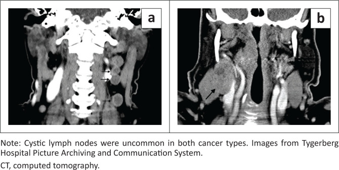
背景:研究发现,与HPV阴性的口咽鳞状细胞癌相比,人乳头瘤病毒(HPV)阳性的口咽鳞状细胞癌(OPSCC)在发病时具有较不晚期的原发肿瘤,更晚期的淋巴结扩散,并且通常具有囊性转移淋巴结。目的:比较南非患者hpv阳性和hpv阴性OPSCC的放射学特征。方法:在南非一家大型医院进行回顾性横断面研究。资格要求在2007年至2023年间具有组织学证明的OPSCC;p16抗原检测,如果阳性,则进行确认性HPV DNA PCR检测和基线治疗前增强颈部CT扫描。纳入了所有符合条件的hpv阳性OPSCC患者和符合条件的hpv阴性OPSCC患者的随机样本。结果:招募了21例hpv阳性和55例hpv阴性的OPSCC患者。在肿瘤中心位置、局部进展(≥T3分别为67%和71%,p = 0.54)、平均原发肿瘤大小(41 mm对39 mm, p = 0.73)、淋巴结扩散(67%对82%,p = 0.22)或形态囊性淋巴结(10%和4%,p = 0.61)方面,两组差异均无统计学意义。结论:在研究的南非患者样本中,hpv阳性和hpv阴性的OPSCC的CT影像学表现无统计学差异。贡献:本研究记录了一小部分南非样本人群中OPSCC的放射学特征,其中hpv阳性和hpv阴性的OPSCC在CT标准上无法区分,并且没有显示文献中描述的经典特征。
The radiological features of HPV-positive vs HPV-negative OPSCC at a South African hospital.
Background: Studies have found that, at presentation, human papillomavirus (HPV)-positive oropharyngeal squamous cell carcinoma (OPSCC) has a less advanced primary tumour, more advanced lymph node spread and commonly has cystic metastatic lymph nodes in comparison to HPV-negative OPSCC.
Objectives: To compare the radiological features of HPV-positive and HPV-negative OPSCC in South African patients.
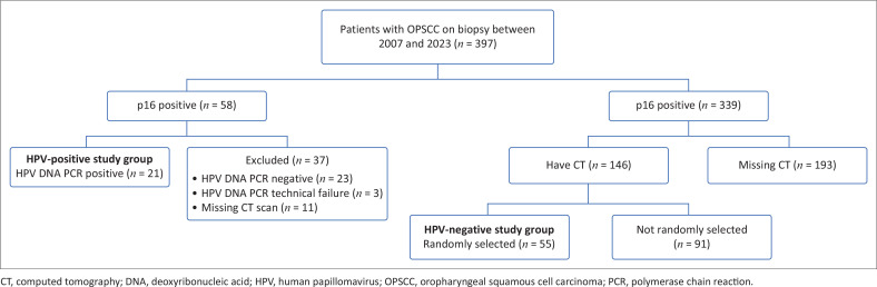
Method: A retrospective cross-sectional study was conducted at a large South African hospital. Eligibility required a histologically proven OPSCC between 2007 and 2023; a p16 antigen test and, if positive, a confirmatory HPV DNA PCR test and a baseline pre-treatment contrast enhanced neck CT scan. All eligible HPV-positive OPSCC patients and a random sample of eligible HPV-negative OPSCC patients were enrolled.
Results: Twenty-one HPV-positive and 55 HPV-negative OPSCC patients were recruited. There was no statistically significant difference in the tumour epicentre location, local advancement (≥ T3 in 67% and 71%, respectively, p = 0.54), mean primary tumour size (41 mm vs. 39 mm, p = 0.73), lymph node spread (bilateral or more in 67% vs. 82%, p = 0.22) or morphologically cystic lymph nodes (10% and 4%, p = 0.61).
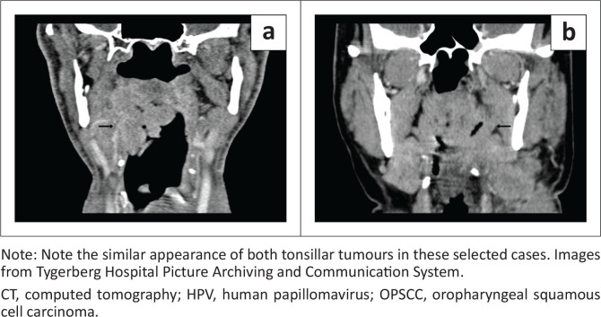
Conclusion: There was no statistically significant difference in the CT imaging appearances of HPV-positive and HPV-negative OPSCC in the studied sample of South African patients.
Contribution: This study documents the radiological features of OPSCC in a small South African sample population, where HPV-positive and HPV-negative OPSCC could not be distinguished on CT criteria and did not display the classic features described in the literature.
期刊介绍:
The SA Journal of Radiology is the official journal of the Radiological Society of South Africa and the Professional Association of Radiologists in South Africa and Namibia. The SA Journal of Radiology is a general diagnostic radiological journal which carries original research and review articles, pictorial essays, case reports, letters, editorials, radiological practice and other radiological articles.

 求助内容:
求助内容: 应助结果提醒方式:


