{"title":"铁人三项运动员自发性纵隔气肿一例","authors":"Jack Golder OMS-II, MS","doi":"10.1002/emp2.13290","DOIUrl":null,"url":null,"abstract":"<p>Spontaneous pneumomediastinum is an infrequent condition typically secondary to smoking, illicit drug use, or asthma. The condition often follows barotrauma or bronchial hyperactivity, causing alveolar destruction and air trapping within the mediastinum. Rarely, it may present following strenuous exercise, particularly in tall, thin males, resembling the presentation of pneumothorax. In this case, a 23-year-old male with no prior medical history presented to the emergency department with chest pain and dyspnea following intense training for a triathlon. Following a normal chest x-ray, a high-resolution computed tomography imaging revealed the presence of spontaneous pneumomediastinum. The patient was admitted for observation and managed conservatively with close monitoring for potential complications such as pneumothorax or pneumopericardium. Emergency physicians should maintain a high index of suspicion for spontaneous pneumomediastinum in patients presenting with acute chest pain and dyspnea, especially in the absence of significant comorbidities. This condition can mimic other cardiopulmonary emergencies, necessitating its inclusion in the differential diagnosis to ensure accurate and timely management.</p>","PeriodicalId":73967,"journal":{"name":"Journal of the American College of Emergency Physicians open","volume":"5 6","pages":""},"PeriodicalIF":1.6000,"publicationDate":"2024-12-04","publicationTypes":"Journal Article","fieldsOfStudy":null,"isOpenAccess":false,"openAccessPdf":"https://onlinelibrary.wiley.com/doi/epdf/10.1002/emp2.13290","citationCount":"0","resultStr":"{\"title\":\"A case of spontaneous pneumomediastinum in a triathlete\",\"authors\":\"Jack Golder OMS-II, MS\",\"doi\":\"10.1002/emp2.13290\",\"DOIUrl\":null,\"url\":null,\"abstract\":\"<p>Spontaneous pneumomediastinum is an infrequent condition typically secondary to smoking, illicit drug use, or asthma. The condition often follows barotrauma or bronchial hyperactivity, causing alveolar destruction and air trapping within the mediastinum. Rarely, it may present following strenuous exercise, particularly in tall, thin males, resembling the presentation of pneumothorax. In this case, a 23-year-old male with no prior medical history presented to the emergency department with chest pain and dyspnea following intense training for a triathlon. Following a normal chest x-ray, a high-resolution computed tomography imaging revealed the presence of spontaneous pneumomediastinum. The patient was admitted for observation and managed conservatively with close monitoring for potential complications such as pneumothorax or pneumopericardium. Emergency physicians should maintain a high index of suspicion for spontaneous pneumomediastinum in patients presenting with acute chest pain and dyspnea, especially in the absence of significant comorbidities. This condition can mimic other cardiopulmonary emergencies, necessitating its inclusion in the differential diagnosis to ensure accurate and timely management.</p>\",\"PeriodicalId\":73967,\"journal\":{\"name\":\"Journal of the American College of Emergency Physicians open\",\"volume\":\"5 6\",\"pages\":\"\"},\"PeriodicalIF\":1.6000,\"publicationDate\":\"2024-12-04\",\"publicationTypes\":\"Journal Article\",\"fieldsOfStudy\":null,\"isOpenAccess\":false,\"openAccessPdf\":\"https://onlinelibrary.wiley.com/doi/epdf/10.1002/emp2.13290\",\"citationCount\":\"0\",\"resultStr\":null,\"platform\":\"Semanticscholar\",\"paperid\":null,\"PeriodicalName\":\"Journal of the American College of Emergency Physicians open\",\"FirstCategoryId\":\"1085\",\"ListUrlMain\":\"https://onlinelibrary.wiley.com/doi/10.1002/emp2.13290\",\"RegionNum\":0,\"RegionCategory\":null,\"ArticlePicture\":[],\"TitleCN\":null,\"AbstractTextCN\":null,\"PMCID\":null,\"EPubDate\":\"\",\"PubModel\":\"\",\"JCR\":\"Q2\",\"JCRName\":\"EMERGENCY MEDICINE\",\"Score\":null,\"Total\":0}","platform":"Semanticscholar","paperid":null,"PeriodicalName":"Journal of the American College of Emergency Physicians open","FirstCategoryId":"1085","ListUrlMain":"https://onlinelibrary.wiley.com/doi/10.1002/emp2.13290","RegionNum":0,"RegionCategory":null,"ArticlePicture":[],"TitleCN":null,"AbstractTextCN":null,"PMCID":null,"EPubDate":"","PubModel":"","JCR":"Q2","JCRName":"EMERGENCY MEDICINE","Score":null,"Total":0}
A case of spontaneous pneumomediastinum in a triathlete
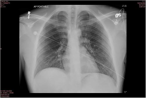
Spontaneous pneumomediastinum is an infrequent condition typically secondary to smoking, illicit drug use, or asthma. The condition often follows barotrauma or bronchial hyperactivity, causing alveolar destruction and air trapping within the mediastinum. Rarely, it may present following strenuous exercise, particularly in tall, thin males, resembling the presentation of pneumothorax. In this case, a 23-year-old male with no prior medical history presented to the emergency department with chest pain and dyspnea following intense training for a triathlon. Following a normal chest x-ray, a high-resolution computed tomography imaging revealed the presence of spontaneous pneumomediastinum. The patient was admitted for observation and managed conservatively with close monitoring for potential complications such as pneumothorax or pneumopericardium. Emergency physicians should maintain a high index of suspicion for spontaneous pneumomediastinum in patients presenting with acute chest pain and dyspnea, especially in the absence of significant comorbidities. This condition can mimic other cardiopulmonary emergencies, necessitating its inclusion in the differential diagnosis to ensure accurate and timely management.

 求助内容:
求助内容: 应助结果提醒方式:


