Qi Ouyang, Fei Zhao, Jingjing Ye, Mengyang Xu, Suyun Pu, Wenxue Hui, Xinyan Gao, Xiaochuan Zha, Hao Chen, Zhiming Wang, Fei Li, Zonghua Luo*, Kurt Wüthrich and Garth J. Thompson*,
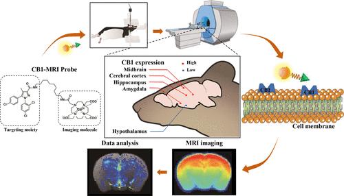
{"title":"Rimota-Gd:用于小鼠脑内大麻素1受体分布的体内MRI研究的顺磁探针","authors":"Qi Ouyang, Fei Zhao, Jingjing Ye, Mengyang Xu, Suyun Pu, Wenxue Hui, Xinyan Gao, Xiaochuan Zha, Hao Chen, Zhiming Wang, Fei Li, Zonghua Luo*, Kurt Wüthrich and Garth J. Thompson*, ","doi":"10.1021/acschemneuro.4c0025910.1021/acschemneuro.4c00259","DOIUrl":null,"url":null,"abstract":"<p >The cannabinoid 1 receptor (CB1) is highly expressed in the central nervous system, where its physiological functions include the regulation of energy balance, pain, and addiction. Herein, we develop and validate a technique to use magnetic resonance imaging (MRI) to investigate the distribution of CB1 across mouse brains with high spatial resolution, expanding previously described in vitro studies and in vivo studies with positron emission tomography (PET). To support the MRI investigations, we developed a ligand that is specific for in vivo neuroimaging of CB1. By chemically conjugating the CB1 antagonist rimonabant acid to a gadolinium chelator, we obtained the paramagnetic probe Rimota-Gd. The specificity of binding of rimonabant acid to CB1 and the relaxation enhancement by the paramagnetic gadolinium permit MRI-based localization of CB1. We used Rimota-Gd to investigate the spatial distribution of CB1 across the mouse brain and compared the results with an investigation using the PET radioligand [<sup>18</sup>F]MK-9470. Rimota-Gd opens the door for in vivo MRI imaging of CB1 and provides a roadmap for the study of other receptors by whole-brain images with high spatial and temporal resolution.</p>","PeriodicalId":13,"journal":{"name":"ACS Chemical Neuroscience","volume":"15 23","pages":"4258–4266 4258–4266"},"PeriodicalIF":3.9000,"publicationDate":"2024-11-14","publicationTypes":"Journal Article","fieldsOfStudy":null,"isOpenAccess":false,"openAccessPdf":"","citationCount":"0","resultStr":"{\"title\":\"Rimota-Gd: Paramagnetic Probe for In Vivo MRI Studies of the Cannabinoid 1 Receptor Distribution in the Mouse Brain\",\"authors\":\"Qi Ouyang, Fei Zhao, Jingjing Ye, Mengyang Xu, Suyun Pu, Wenxue Hui, Xinyan Gao, Xiaochuan Zha, Hao Chen, Zhiming Wang, Fei Li, Zonghua Luo*, Kurt Wüthrich and Garth J. Thompson*, \",\"doi\":\"10.1021/acschemneuro.4c0025910.1021/acschemneuro.4c00259\",\"DOIUrl\":null,\"url\":null,\"abstract\":\"<p >The cannabinoid 1 receptor (CB1) is highly expressed in the central nervous system, where its physiological functions include the regulation of energy balance, pain, and addiction. Herein, we develop and validate a technique to use magnetic resonance imaging (MRI) to investigate the distribution of CB1 across mouse brains with high spatial resolution, expanding previously described in vitro studies and in vivo studies with positron emission tomography (PET). To support the MRI investigations, we developed a ligand that is specific for in vivo neuroimaging of CB1. By chemically conjugating the CB1 antagonist rimonabant acid to a gadolinium chelator, we obtained the paramagnetic probe Rimota-Gd. The specificity of binding of rimonabant acid to CB1 and the relaxation enhancement by the paramagnetic gadolinium permit MRI-based localization of CB1. We used Rimota-Gd to investigate the spatial distribution of CB1 across the mouse brain and compared the results with an investigation using the PET radioligand [<sup>18</sup>F]MK-9470. Rimota-Gd opens the door for in vivo MRI imaging of CB1 and provides a roadmap for the study of other receptors by whole-brain images with high spatial and temporal resolution.</p>\",\"PeriodicalId\":13,\"journal\":{\"name\":\"ACS Chemical Neuroscience\",\"volume\":\"15 23\",\"pages\":\"4258–4266 4258–4266\"},\"PeriodicalIF\":3.9000,\"publicationDate\":\"2024-11-14\",\"publicationTypes\":\"Journal Article\",\"fieldsOfStudy\":null,\"isOpenAccess\":false,\"openAccessPdf\":\"\",\"citationCount\":\"0\",\"resultStr\":null,\"platform\":\"Semanticscholar\",\"paperid\":null,\"PeriodicalName\":\"ACS Chemical Neuroscience\",\"FirstCategoryId\":\"3\",\"ListUrlMain\":\"https://pubs.acs.org/doi/10.1021/acschemneuro.4c00259\",\"RegionNum\":3,\"RegionCategory\":\"医学\",\"ArticlePicture\":[],\"TitleCN\":null,\"AbstractTextCN\":null,\"PMCID\":null,\"EPubDate\":\"\",\"PubModel\":\"\",\"JCR\":\"Q2\",\"JCRName\":\"BIOCHEMISTRY & MOLECULAR BIOLOGY\",\"Score\":null,\"Total\":0}","platform":"Semanticscholar","paperid":null,"PeriodicalName":"ACS Chemical Neuroscience","FirstCategoryId":"3","ListUrlMain":"https://pubs.acs.org/doi/10.1021/acschemneuro.4c00259","RegionNum":3,"RegionCategory":"医学","ArticlePicture":[],"TitleCN":null,"AbstractTextCN":null,"PMCID":null,"EPubDate":"","PubModel":"","JCR":"Q2","JCRName":"BIOCHEMISTRY & MOLECULAR BIOLOGY","Score":null,"Total":0}
Rimota-Gd: Paramagnetic Probe for In Vivo MRI Studies of the Cannabinoid 1 Receptor Distribution in the Mouse Brain
The cannabinoid 1 receptor (CB1) is highly expressed in the central nervous system, where its physiological functions include the regulation of energy balance, pain, and addiction. Herein, we develop and validate a technique to use magnetic resonance imaging (MRI) to investigate the distribution of CB1 across mouse brains with high spatial resolution, expanding previously described in vitro studies and in vivo studies with positron emission tomography (PET). To support the MRI investigations, we developed a ligand that is specific for in vivo neuroimaging of CB1. By chemically conjugating the CB1 antagonist rimonabant acid to a gadolinium chelator, we obtained the paramagnetic probe Rimota-Gd. The specificity of binding of rimonabant acid to CB1 and the relaxation enhancement by the paramagnetic gadolinium permit MRI-based localization of CB1. We used Rimota-Gd to investigate the spatial distribution of CB1 across the mouse brain and compared the results with an investigation using the PET radioligand [18F]MK-9470. Rimota-Gd opens the door for in vivo MRI imaging of CB1 and provides a roadmap for the study of other receptors by whole-brain images with high spatial and temporal resolution.
期刊介绍:
ACS Chemical Neuroscience publishes high-quality research articles and reviews that showcase chemical, quantitative biological, biophysical and bioengineering approaches to the understanding of the nervous system and to the development of new treatments for neurological disorders. Research in the journal focuses on aspects of chemical neurobiology and bio-neurochemistry such as the following:
Neurotransmitters and receptors
Neuropharmaceuticals and therapeutics
Neural development—Plasticity, and degeneration
Chemical, physical, and computational methods in neuroscience
Neuronal diseases—basis, detection, and treatment
Mechanism of aging, learning, memory and behavior
Pain and sensory processing
Neurotoxins
Neuroscience-inspired bioengineering
Development of methods in chemical neurobiology
Neuroimaging agents and technologies
Animal models for central nervous system diseases
Behavioral research
 求助内容:
求助内容: 应助结果提醒方式:


