Louis Lassalle, Nor-Eddine Regnard, Marion Durteste, Jeanne Ventre, Vincent Marty, Lauryane Clovis, Zekun Zhang, Nicolas Nitche, Alexis Ducarouge, Jean-Denis Laredo, Ali Guermazi
{"title":"对全腿站立式x光片自动测量的深度学习软件的评估。","authors":"Louis Lassalle, Nor-Eddine Regnard, Marion Durteste, Jeanne Ventre, Vincent Marty, Lauryane Clovis, Zekun Zhang, Nicolas Nitche, Alexis Ducarouge, Jean-Denis Laredo, Ali Guermazi","doi":"10.1186/s43019-024-00246-1","DOIUrl":null,"url":null,"abstract":"<p><strong>Background: </strong>Precise lower limb measurements are crucial for assessing musculoskeletal health; fully automated solutions have the potential to enhance standardization and reproducibility of these measurements. This study compared the measurements performed by BoneMetrics (Gleamer, Paris, France), a commercial artificial intelligence (AI)-based software, to expert manual measurements on anteroposterior full-leg standing radiographs.</p><p><strong>Methods: </strong>A retrospective analysis was conducted on a dataset comprising consecutive anteroposterior full-leg standing radiographs obtained from four imaging institutions. Key anatomical landmarks to define the hip-knee-ankle angle, pelvic obliquity, leg length, femoral length, and tibial length were annotated independently by two expert musculoskeletal radiologists and served as the ground truth. The performance of the AI was compared against these reference measurements using the mean absolute error, Bland-Altman analyses, and intraclass correlation coefficients.</p><p><strong>Results: </strong>A total of 175 anteroposterior full-leg standing radiographs from 167 patients were included in the final dataset (mean age = 49.9 ± 23.6 years old; 103 women and 64 men). Mean absolute error values were 0.30° (95% confidence interval [CI] [0.28, 0.32]) for the hip-knee-ankle angle, 0.75 mm (95% CI [0.60, 0.88]) for pelvic obliquity, 1.03 mm (95% CI [0.91,1.14]) for leg length from the top of the femoral head, 1.45 mm (95% CI [1.33, 1.60]) for leg length from the center of the femoral head, 0.95 mm (95% CI [0.85, 1.04]) for femoral length from the top of the femoral head, 1.23 mm (95% CI [1.12, 1.32]) for femoral length from the center of the femoral head, and 1.38 mm (95% CI [1.21, 1.52]) for tibial length. The Bland-Altman analyses revealed no systematic bias across all measurements. Additionally, the software exhibited excellent agreement with the gold-standard measurements with intraclass correlation coefficient (ICC) values above 0.97 for all parameters.</p><p><strong>Conclusions: </strong>Automated measurements on anteroposterior full-leg standing radiographs offer a reliable alternative to manual assessments. The use of AI in musculoskeletal radiology has the potential to support physicians in their daily practice without compromising patient care standards.</p>","PeriodicalId":36317,"journal":{"name":"Knee Surgery and Related Research","volume":"36 1","pages":"40"},"PeriodicalIF":4.4000,"publicationDate":"2024-11-29","publicationTypes":"Journal Article","fieldsOfStudy":null,"isOpenAccess":false,"openAccessPdf":"https://www.ncbi.nlm.nih.gov/pmc/articles/PMC11606017/pdf/","citationCount":"0","resultStr":"{\"title\":\"Evaluation of a deep learning software for automated measurements on full-leg standing radiographs.\",\"authors\":\"Louis Lassalle, Nor-Eddine Regnard, Marion Durteste, Jeanne Ventre, Vincent Marty, Lauryane Clovis, Zekun Zhang, Nicolas Nitche, Alexis Ducarouge, Jean-Denis Laredo, Ali Guermazi\",\"doi\":\"10.1186/s43019-024-00246-1\",\"DOIUrl\":null,\"url\":null,\"abstract\":\"<p><strong>Background: </strong>Precise lower limb measurements are crucial for assessing musculoskeletal health; fully automated solutions have the potential to enhance standardization and reproducibility of these measurements. This study compared the measurements performed by BoneMetrics (Gleamer, Paris, France), a commercial artificial intelligence (AI)-based software, to expert manual measurements on anteroposterior full-leg standing radiographs.</p><p><strong>Methods: </strong>A retrospective analysis was conducted on a dataset comprising consecutive anteroposterior full-leg standing radiographs obtained from four imaging institutions. Key anatomical landmarks to define the hip-knee-ankle angle, pelvic obliquity, leg length, femoral length, and tibial length were annotated independently by two expert musculoskeletal radiologists and served as the ground truth. The performance of the AI was compared against these reference measurements using the mean absolute error, Bland-Altman analyses, and intraclass correlation coefficients.</p><p><strong>Results: </strong>A total of 175 anteroposterior full-leg standing radiographs from 167 patients were included in the final dataset (mean age = 49.9 ± 23.6 years old; 103 women and 64 men). Mean absolute error values were 0.30° (95% confidence interval [CI] [0.28, 0.32]) for the hip-knee-ankle angle, 0.75 mm (95% CI [0.60, 0.88]) for pelvic obliquity, 1.03 mm (95% CI [0.91,1.14]) for leg length from the top of the femoral head, 1.45 mm (95% CI [1.33, 1.60]) for leg length from the center of the femoral head, 0.95 mm (95% CI [0.85, 1.04]) for femoral length from the top of the femoral head, 1.23 mm (95% CI [1.12, 1.32]) for femoral length from the center of the femoral head, and 1.38 mm (95% CI [1.21, 1.52]) for tibial length. The Bland-Altman analyses revealed no systematic bias across all measurements. Additionally, the software exhibited excellent agreement with the gold-standard measurements with intraclass correlation coefficient (ICC) values above 0.97 for all parameters.</p><p><strong>Conclusions: </strong>Automated measurements on anteroposterior full-leg standing radiographs offer a reliable alternative to manual assessments. The use of AI in musculoskeletal radiology has the potential to support physicians in their daily practice without compromising patient care standards.</p>\",\"PeriodicalId\":36317,\"journal\":{\"name\":\"Knee Surgery and Related Research\",\"volume\":\"36 1\",\"pages\":\"40\"},\"PeriodicalIF\":4.4000,\"publicationDate\":\"2024-11-29\",\"publicationTypes\":\"Journal Article\",\"fieldsOfStudy\":null,\"isOpenAccess\":false,\"openAccessPdf\":\"https://www.ncbi.nlm.nih.gov/pmc/articles/PMC11606017/pdf/\",\"citationCount\":\"0\",\"resultStr\":null,\"platform\":\"Semanticscholar\",\"paperid\":null,\"PeriodicalName\":\"Knee Surgery and Related Research\",\"FirstCategoryId\":\"1085\",\"ListUrlMain\":\"https://doi.org/10.1186/s43019-024-00246-1\",\"RegionNum\":0,\"RegionCategory\":null,\"ArticlePicture\":[],\"TitleCN\":null,\"AbstractTextCN\":null,\"PMCID\":null,\"EPubDate\":\"\",\"PubModel\":\"\",\"JCR\":\"Q2\",\"JCRName\":\"Medicine\",\"Score\":null,\"Total\":0}","platform":"Semanticscholar","paperid":null,"PeriodicalName":"Knee Surgery and Related Research","FirstCategoryId":"1085","ListUrlMain":"https://doi.org/10.1186/s43019-024-00246-1","RegionNum":0,"RegionCategory":null,"ArticlePicture":[],"TitleCN":null,"AbstractTextCN":null,"PMCID":null,"EPubDate":"","PubModel":"","JCR":"Q2","JCRName":"Medicine","Score":null,"Total":0}
Evaluation of a deep learning software for automated measurements on full-leg standing radiographs.
Background: Precise lower limb measurements are crucial for assessing musculoskeletal health; fully automated solutions have the potential to enhance standardization and reproducibility of these measurements. This study compared the measurements performed by BoneMetrics (Gleamer, Paris, France), a commercial artificial intelligence (AI)-based software, to expert manual measurements on anteroposterior full-leg standing radiographs.
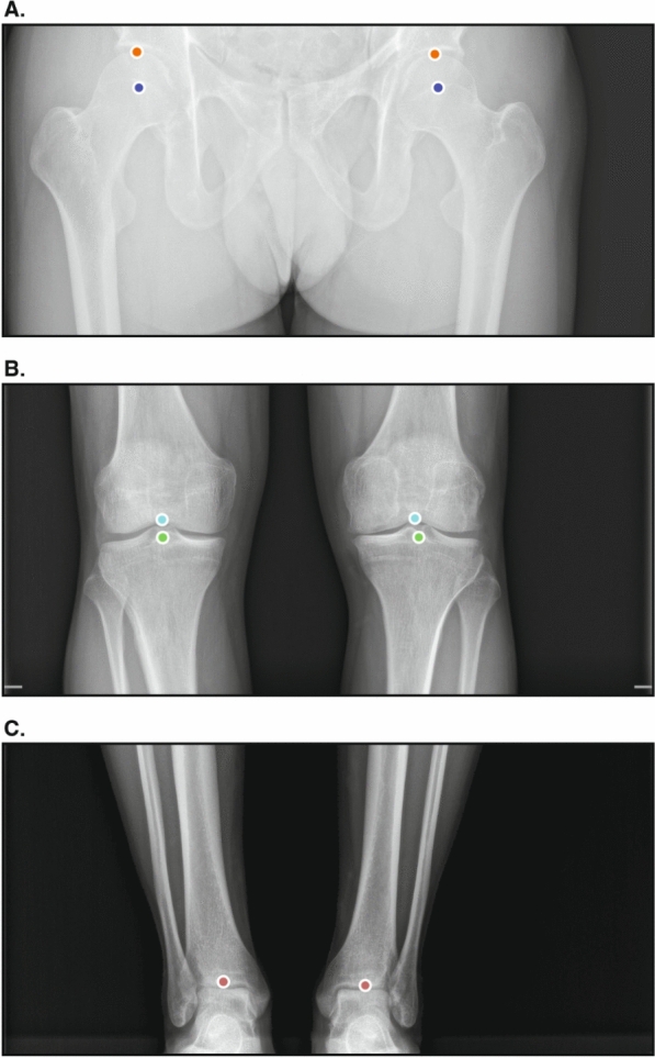
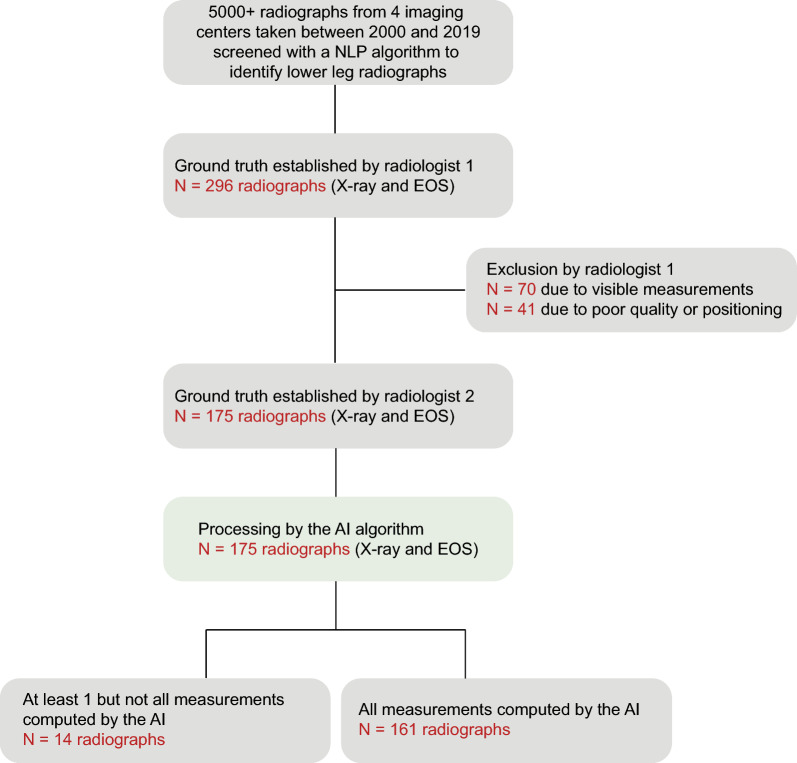
Methods: A retrospective analysis was conducted on a dataset comprising consecutive anteroposterior full-leg standing radiographs obtained from four imaging institutions. Key anatomical landmarks to define the hip-knee-ankle angle, pelvic obliquity, leg length, femoral length, and tibial length were annotated independently by two expert musculoskeletal radiologists and served as the ground truth. The performance of the AI was compared against these reference measurements using the mean absolute error, Bland-Altman analyses, and intraclass correlation coefficients.
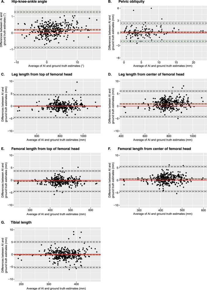
Results: A total of 175 anteroposterior full-leg standing radiographs from 167 patients were included in the final dataset (mean age = 49.9 ± 23.6 years old; 103 women and 64 men). Mean absolute error values were 0.30° (95% confidence interval [CI] [0.28, 0.32]) for the hip-knee-ankle angle, 0.75 mm (95% CI [0.60, 0.88]) for pelvic obliquity, 1.03 mm (95% CI [0.91,1.14]) for leg length from the top of the femoral head, 1.45 mm (95% CI [1.33, 1.60]) for leg length from the center of the femoral head, 0.95 mm (95% CI [0.85, 1.04]) for femoral length from the top of the femoral head, 1.23 mm (95% CI [1.12, 1.32]) for femoral length from the center of the femoral head, and 1.38 mm (95% CI [1.21, 1.52]) for tibial length. The Bland-Altman analyses revealed no systematic bias across all measurements. Additionally, the software exhibited excellent agreement with the gold-standard measurements with intraclass correlation coefficient (ICC) values above 0.97 for all parameters.
Conclusions: Automated measurements on anteroposterior full-leg standing radiographs offer a reliable alternative to manual assessments. The use of AI in musculoskeletal radiology has the potential to support physicians in their daily practice without compromising patient care standards.

 求助内容:
求助内容: 应助结果提醒方式:


