Alessandro Fontanella, Wenwen Li, Grant Mair, Antreas Antoniou, Eleanor Platt, Paul Armitage, Emanuele Trucco, Joanna M Wardlaw, Amos Storkey
{"title":"开发一种深度学习方法来识别脑 CT 上的急性缺血性中风病灶。","authors":"Alessandro Fontanella, Wenwen Li, Grant Mair, Antreas Antoniou, Eleanor Platt, Paul Armitage, Emanuele Trucco, Joanna M Wardlaw, Amos Storkey","doi":"10.1136/svn-2024-003372","DOIUrl":null,"url":null,"abstract":"<p><strong>Background: </strong>CT is commonly used to image patients with ischaemic stroke but radiologist interpretation may be delayed. Machine learning techniques can provide rapid automated CT assessment but are usually developed from annotated images which necessarily limits the size and representation of development data sets. We aimed to develop a deep learning (DL) method using CT brain scans that were labelled but not annotated for the presence of ischaemic lesions.</p><p><strong>Methods: </strong>We designed a convolutional neural network-based DL algorithm to detect ischaemic lesions on CT. Our algorithm was trained using routinely acquired CT brain scans collected for a large multicentre international trial. These scans had previously been labelled by experts for acute and chronic appearances. We explored the impact of ischaemic lesion features, background brain appearances and timing of CT (baseline or 24-48 hour follow-up) on DL performance.</p><p><strong>Results: </strong>From 5772 CT scans of 2347 patients (median age 82), 54% had visible ischaemic lesions according to experts. Our DL method achieved 72% accuracy in detecting ischaemic lesions. Detection was better for larger (80% accuracy) or multiple (87% accuracy for two, 100% for three or more) lesions and with follow-up scans (76% accuracy vs 67% at baseline). Chronic brain conditions reduced accuracy, particularly non-stroke lesions and old stroke lesions (32% and 31% error rates, respectively).</p><p><strong>Conclusion: </strong>DL methods can be designed for ischaemic lesion detection on CT using the vast quantities of routinely collected brain scans without the need for lesion annotation. Ultimately, this should lead to more robust and widely applicable methods.</p>","PeriodicalId":48733,"journal":{"name":"Journal of Investigative Medicine","volume":" ","pages":"499-507"},"PeriodicalIF":4.9000,"publicationDate":"2025-08-26","publicationTypes":"Journal Article","fieldsOfStudy":null,"isOpenAccess":false,"openAccessPdf":"https://www.ncbi.nlm.nih.gov/pmc/articles/PMC12415648/pdf/","citationCount":"0","resultStr":"{\"title\":\"Development of a deep learning method to identify acute ischaemic stroke lesions on brain CT.\",\"authors\":\"Alessandro Fontanella, Wenwen Li, Grant Mair, Antreas Antoniou, Eleanor Platt, Paul Armitage, Emanuele Trucco, Joanna M Wardlaw, Amos Storkey\",\"doi\":\"10.1136/svn-2024-003372\",\"DOIUrl\":null,\"url\":null,\"abstract\":\"<p><strong>Background: </strong>CT is commonly used to image patients with ischaemic stroke but radiologist interpretation may be delayed. Machine learning techniques can provide rapid automated CT assessment but are usually developed from annotated images which necessarily limits the size and representation of development data sets. We aimed to develop a deep learning (DL) method using CT brain scans that were labelled but not annotated for the presence of ischaemic lesions.</p><p><strong>Methods: </strong>We designed a convolutional neural network-based DL algorithm to detect ischaemic lesions on CT. Our algorithm was trained using routinely acquired CT brain scans collected for a large multicentre international trial. These scans had previously been labelled by experts for acute and chronic appearances. We explored the impact of ischaemic lesion features, background brain appearances and timing of CT (baseline or 24-48 hour follow-up) on DL performance.</p><p><strong>Results: </strong>From 5772 CT scans of 2347 patients (median age 82), 54% had visible ischaemic lesions according to experts. Our DL method achieved 72% accuracy in detecting ischaemic lesions. Detection was better for larger (80% accuracy) or multiple (87% accuracy for two, 100% for three or more) lesions and with follow-up scans (76% accuracy vs 67% at baseline). Chronic brain conditions reduced accuracy, particularly non-stroke lesions and old stroke lesions (32% and 31% error rates, respectively).</p><p><strong>Conclusion: </strong>DL methods can be designed for ischaemic lesion detection on CT using the vast quantities of routinely collected brain scans without the need for lesion annotation. Ultimately, this should lead to more robust and widely applicable methods.</p>\",\"PeriodicalId\":48733,\"journal\":{\"name\":\"Journal of Investigative Medicine\",\"volume\":\" \",\"pages\":\"499-507\"},\"PeriodicalIF\":4.9000,\"publicationDate\":\"2025-08-26\",\"publicationTypes\":\"Journal Article\",\"fieldsOfStudy\":null,\"isOpenAccess\":false,\"openAccessPdf\":\"https://www.ncbi.nlm.nih.gov/pmc/articles/PMC12415648/pdf/\",\"citationCount\":\"0\",\"resultStr\":null,\"platform\":\"Semanticscholar\",\"paperid\":null,\"PeriodicalName\":\"Journal of Investigative Medicine\",\"FirstCategoryId\":\"3\",\"ListUrlMain\":\"https://doi.org/10.1136/svn-2024-003372\",\"RegionNum\":1,\"RegionCategory\":\"医学\",\"ArticlePicture\":[],\"TitleCN\":null,\"AbstractTextCN\":null,\"PMCID\":null,\"EPubDate\":\"\",\"PubModel\":\"\",\"JCR\":\"\",\"JCRName\":\"\",\"Score\":null,\"Total\":0}","platform":"Semanticscholar","paperid":null,"PeriodicalName":"Journal of Investigative Medicine","FirstCategoryId":"3","ListUrlMain":"https://doi.org/10.1136/svn-2024-003372","RegionNum":1,"RegionCategory":"医学","ArticlePicture":[],"TitleCN":null,"AbstractTextCN":null,"PMCID":null,"EPubDate":"","PubModel":"","JCR":"","JCRName":"","Score":null,"Total":0}
Development of a deep learning method to identify acute ischaemic stroke lesions on brain CT.
Background: CT is commonly used to image patients with ischaemic stroke but radiologist interpretation may be delayed. Machine learning techniques can provide rapid automated CT assessment but are usually developed from annotated images which necessarily limits the size and representation of development data sets. We aimed to develop a deep learning (DL) method using CT brain scans that were labelled but not annotated for the presence of ischaemic lesions.
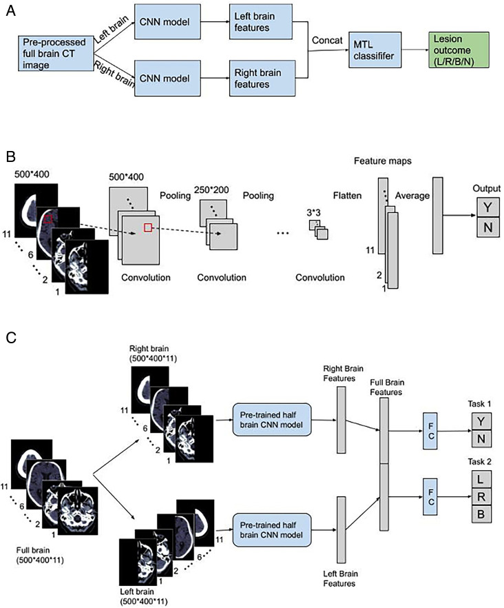
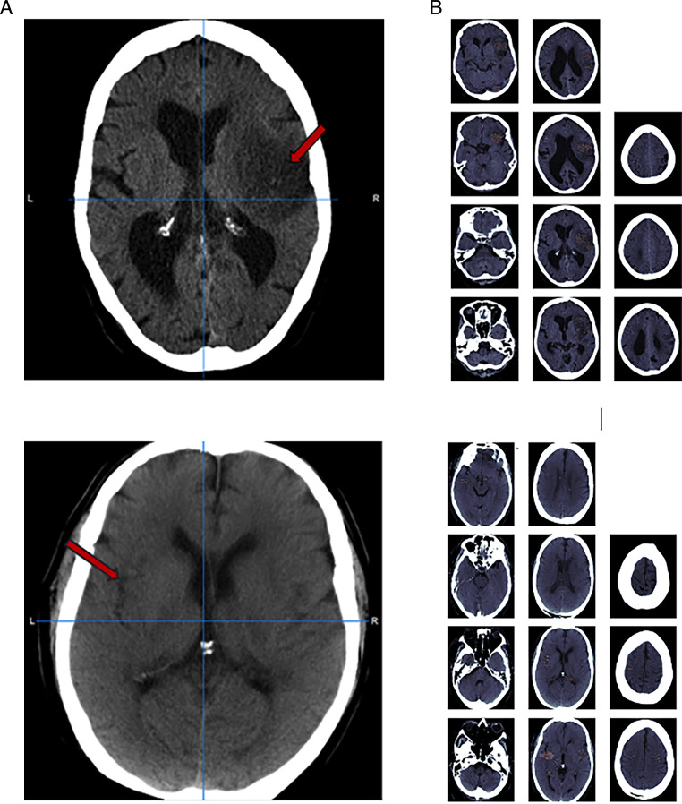
Methods: We designed a convolutional neural network-based DL algorithm to detect ischaemic lesions on CT. Our algorithm was trained using routinely acquired CT brain scans collected for a large multicentre international trial. These scans had previously been labelled by experts for acute and chronic appearances. We explored the impact of ischaemic lesion features, background brain appearances and timing of CT (baseline or 24-48 hour follow-up) on DL performance.
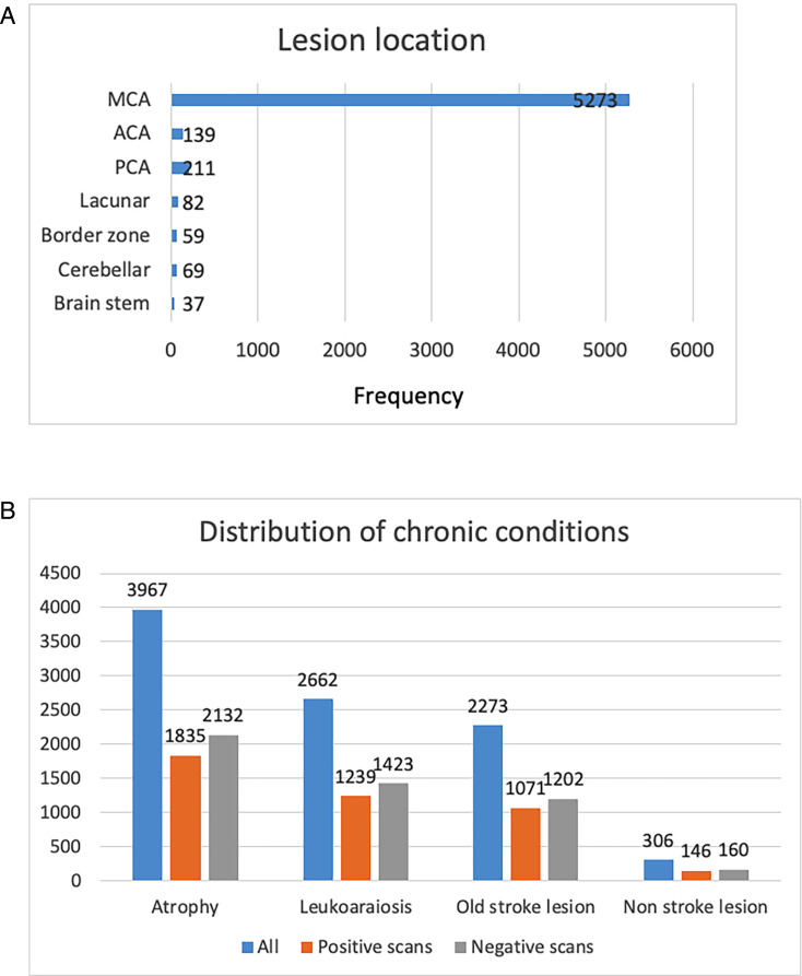
Results: From 5772 CT scans of 2347 patients (median age 82), 54% had visible ischaemic lesions according to experts. Our DL method achieved 72% accuracy in detecting ischaemic lesions. Detection was better for larger (80% accuracy) or multiple (87% accuracy for two, 100% for three or more) lesions and with follow-up scans (76% accuracy vs 67% at baseline). Chronic brain conditions reduced accuracy, particularly non-stroke lesions and old stroke lesions (32% and 31% error rates, respectively).
Conclusion: DL methods can be designed for ischaemic lesion detection on CT using the vast quantities of routinely collected brain scans without the need for lesion annotation. Ultimately, this should lead to more robust and widely applicable methods.
期刊介绍:
Journal of Investigative Medicine (JIM) is the official publication of the American Federation for Medical Research. The journal is peer-reviewed and publishes high-quality original articles and reviews in the areas of basic, clinical, and translational medical research.
JIM publishes on all topics and specialty areas that are critical to the conduct of the entire spectrum of biomedical research: from the translation of clinical observations at the bedside, to basic and animal research to clinical research and the implementation of innovative medical care.
 求助内容:
求助内容: 应助结果提醒方式:


