Ruben D Houvast, Maurice van Duijvenvoorde, Kira Thijse, Wobbe O de Steur, Lioe-Fee de Geus-Oei, A Stijn L P Crobach, Jacobus Burggraaf, Alexander L Vahrmeijer, Peter J K Kuppen
{"title":"选择胃癌分子成像的靶点:免疫组化评估","authors":"Ruben D Houvast, Maurice van Duijvenvoorde, Kira Thijse, Wobbe O de Steur, Lioe-Fee de Geus-Oei, A Stijn L P Crobach, Jacobus Burggraaf, Alexander L Vahrmeijer, Peter J K Kuppen","doi":"10.1007/s40291-024-00755-5","DOIUrl":null,"url":null,"abstract":"<p><strong>Purpose: </strong>Tumor-targeted positron emission tomography (PET) and fluorescence-guided surgery (FGS) could address current challenges in pre- and intraoperative imaging of gastric cancer. Adequate selection of molecular imaging targets remains crucial for successful tumor visualization. This study evaluated the potential of integrin α<sub>v</sub>β<sub>6</sub>, carcinoembryonic antigen-related cell adhesion molecule 5 (CEACAM5), epidermal growth factor receptor (EGFR), epithelial cell adhesion molecule (EpCAM) and human epidermal growth factor receptor-2 (HER2) for molecular imaging of primary gastric cancer, as well as lymph node and distant metastases.</p><p><strong>Methods: </strong>Expression of α<sub>v</sub>β<sub>6</sub>, CEACAM5, EGFR, EpCAM and HER2 was determined using immunohistochemistry in human tissue specimens of primary gastric adenocarcinoma, healthy surrounding stomach, esophageal and duodenal tissue, tumor-positive and tumor-negative lymph nodes, and distant metastases, followed by quantification using the total immunostaining score (TIS).</p><p><strong>Results: </strong>Positive biomarker expression in primary gastric tumors was observed in 86% for α<sub>v</sub>β<sub>6</sub>, 72% for CEACAM5, 77% for EGFR, 93% for EpCAM and 71% for HER2. Tumor expression of CEACAM5, EGFR and EpCAM was higher compared to healthy stomach tissue expression, while this was not the case for α<sub>v</sub>β<sub>6</sub> and HER2. Tumor-positive lymph nodes could be distinguished from tumor-negative lymph nodes, with accuracy ranging from 82 to 93% between biomarkers. CEACAM5, EGFR and EpCAM were abundantly expressed on distant metastases, with expression in 88-95% of tissue specimens.</p><p><strong>Conclusion: </strong>Our findings show that CEACAM5, EGFR and EpCAM are promising targets for molecular imaging of primary gastric cancer, as well as visualization of both lymph node and distant metastases. Further clinical evaluation of PET and FGS tracers targeting these antigens is warranted.</p>","PeriodicalId":49797,"journal":{"name":"Molecular Diagnosis & Therapy","volume":" ","pages":"213-227"},"PeriodicalIF":4.4000,"publicationDate":"2025-03-01","publicationTypes":"Journal Article","fieldsOfStudy":null,"isOpenAccess":false,"openAccessPdf":"https://www.ncbi.nlm.nih.gov/pmc/articles/PMC11860997/pdf/","citationCount":"0","resultStr":"{\"title\":\"Selecting Targets for Molecular Imaging of Gastric Cancer: An Immunohistochemical Evaluation.\",\"authors\":\"Ruben D Houvast, Maurice van Duijvenvoorde, Kira Thijse, Wobbe O de Steur, Lioe-Fee de Geus-Oei, A Stijn L P Crobach, Jacobus Burggraaf, Alexander L Vahrmeijer, Peter J K Kuppen\",\"doi\":\"10.1007/s40291-024-00755-5\",\"DOIUrl\":null,\"url\":null,\"abstract\":\"<p><strong>Purpose: </strong>Tumor-targeted positron emission tomography (PET) and fluorescence-guided surgery (FGS) could address current challenges in pre- and intraoperative imaging of gastric cancer. Adequate selection of molecular imaging targets remains crucial for successful tumor visualization. This study evaluated the potential of integrin α<sub>v</sub>β<sub>6</sub>, carcinoembryonic antigen-related cell adhesion molecule 5 (CEACAM5), epidermal growth factor receptor (EGFR), epithelial cell adhesion molecule (EpCAM) and human epidermal growth factor receptor-2 (HER2) for molecular imaging of primary gastric cancer, as well as lymph node and distant metastases.</p><p><strong>Methods: </strong>Expression of α<sub>v</sub>β<sub>6</sub>, CEACAM5, EGFR, EpCAM and HER2 was determined using immunohistochemistry in human tissue specimens of primary gastric adenocarcinoma, healthy surrounding stomach, esophageal and duodenal tissue, tumor-positive and tumor-negative lymph nodes, and distant metastases, followed by quantification using the total immunostaining score (TIS).</p><p><strong>Results: </strong>Positive biomarker expression in primary gastric tumors was observed in 86% for α<sub>v</sub>β<sub>6</sub>, 72% for CEACAM5, 77% for EGFR, 93% for EpCAM and 71% for HER2. Tumor expression of CEACAM5, EGFR and EpCAM was higher compared to healthy stomach tissue expression, while this was not the case for α<sub>v</sub>β<sub>6</sub> and HER2. Tumor-positive lymph nodes could be distinguished from tumor-negative lymph nodes, with accuracy ranging from 82 to 93% between biomarkers. CEACAM5, EGFR and EpCAM were abundantly expressed on distant metastases, with expression in 88-95% of tissue specimens.</p><p><strong>Conclusion: </strong>Our findings show that CEACAM5, EGFR and EpCAM are promising targets for molecular imaging of primary gastric cancer, as well as visualization of both lymph node and distant metastases. Further clinical evaluation of PET and FGS tracers targeting these antigens is warranted.</p>\",\"PeriodicalId\":49797,\"journal\":{\"name\":\"Molecular Diagnosis & Therapy\",\"volume\":\" \",\"pages\":\"213-227\"},\"PeriodicalIF\":4.4000,\"publicationDate\":\"2025-03-01\",\"publicationTypes\":\"Journal Article\",\"fieldsOfStudy\":null,\"isOpenAccess\":false,\"openAccessPdf\":\"https://www.ncbi.nlm.nih.gov/pmc/articles/PMC11860997/pdf/\",\"citationCount\":\"0\",\"resultStr\":null,\"platform\":\"Semanticscholar\",\"paperid\":null,\"PeriodicalName\":\"Molecular Diagnosis & Therapy\",\"FirstCategoryId\":\"3\",\"ListUrlMain\":\"https://doi.org/10.1007/s40291-024-00755-5\",\"RegionNum\":3,\"RegionCategory\":\"医学\",\"ArticlePicture\":[],\"TitleCN\":null,\"AbstractTextCN\":null,\"PMCID\":null,\"EPubDate\":\"2024/11/14 0:00:00\",\"PubModel\":\"Epub\",\"JCR\":\"Q1\",\"JCRName\":\"GENETICS & HEREDITY\",\"Score\":null,\"Total\":0}","platform":"Semanticscholar","paperid":null,"PeriodicalName":"Molecular Diagnosis & Therapy","FirstCategoryId":"3","ListUrlMain":"https://doi.org/10.1007/s40291-024-00755-5","RegionNum":3,"RegionCategory":"医学","ArticlePicture":[],"TitleCN":null,"AbstractTextCN":null,"PMCID":null,"EPubDate":"2024/11/14 0:00:00","PubModel":"Epub","JCR":"Q1","JCRName":"GENETICS & HEREDITY","Score":null,"Total":0}
引用次数: 0
摘要
目的:肿瘤靶向正电子发射断层扫描(PET)和荧光引导手术(FGS)可解决目前胃癌术前和术中成像的难题。充分选择分子成像靶点仍是成功实现肿瘤可视化的关键。本研究评估了整合素αvβ6、癌胚抗原相关细胞粘附分子5(CEACAM5)、表皮生长因子受体(EGFR)、上皮细胞粘附分子(EpCAM)和人表皮生长因子受体-2(HER2)在原发性胃癌以及淋巴结和远处转移灶分子成像中的潜力:方法:采用免疫组化方法测定原发性胃腺癌、周围健康的胃、食管和十二指肠组织、肿瘤阳性和肿瘤阴性淋巴结以及远处转移灶的人体组织标本中αvβ6、CEACAM5、表皮生长因子受体、EpCAM和HER2的表达,然后用免疫染色总评分(TIS)进行量化:结果:原发性胃肿瘤中αvβ6、CEACAM5、EGFR、EpCAM和HER2的生物标记物阳性表达率分别为86%、72%、77%、93%和71%。CEACAM5、表皮生长因子受体(EGFR)和EpCAM的肿瘤表达高于健康胃组织的表达,而αvβ6和HER2的肿瘤表达则不同。肿瘤阳性淋巴结与肿瘤阴性淋巴结可以区分,生物标记物之间的准确率在82%到93%之间。CEACAM5、表皮生长因子受体(EGFR)和表皮生长因子受体(EpCAM)在远处转移灶中大量表达,88%-95%的组织标本中都有表达:我们的研究结果表明,CEACAM5、表皮生长因子受体(EGFR)和 EpCAM 是原发性胃癌分子成像以及淋巴结和远处转移灶可视化的理想靶点。针对这些抗原的 PET 和 FGS 示踪剂需要进一步的临床评估。
Selecting Targets for Molecular Imaging of Gastric Cancer: An Immunohistochemical Evaluation.
Purpose: Tumor-targeted positron emission tomography (PET) and fluorescence-guided surgery (FGS) could address current challenges in pre- and intraoperative imaging of gastric cancer. Adequate selection of molecular imaging targets remains crucial for successful tumor visualization. This study evaluated the potential of integrin αvβ6, carcinoembryonic antigen-related cell adhesion molecule 5 (CEACAM5), epidermal growth factor receptor (EGFR), epithelial cell adhesion molecule (EpCAM) and human epidermal growth factor receptor-2 (HER2) for molecular imaging of primary gastric cancer, as well as lymph node and distant metastases.
Methods: Expression of αvβ6, CEACAM5, EGFR, EpCAM and HER2 was determined using immunohistochemistry in human tissue specimens of primary gastric adenocarcinoma, healthy surrounding stomach, esophageal and duodenal tissue, tumor-positive and tumor-negative lymph nodes, and distant metastases, followed by quantification using the total immunostaining score (TIS).
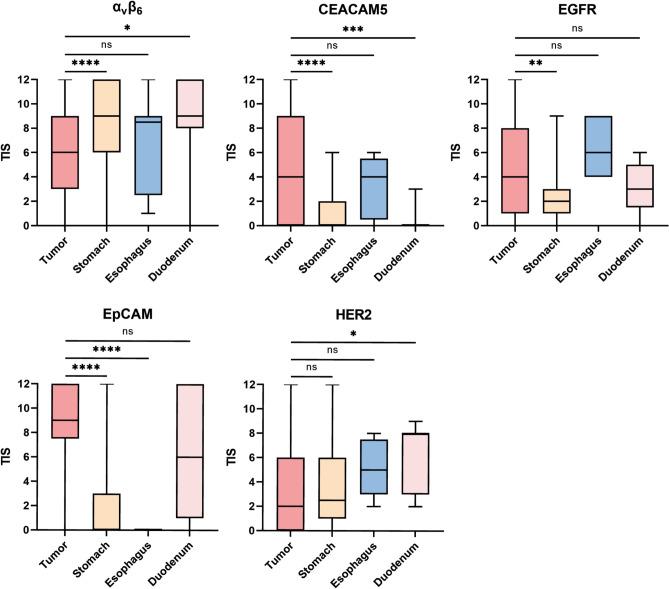
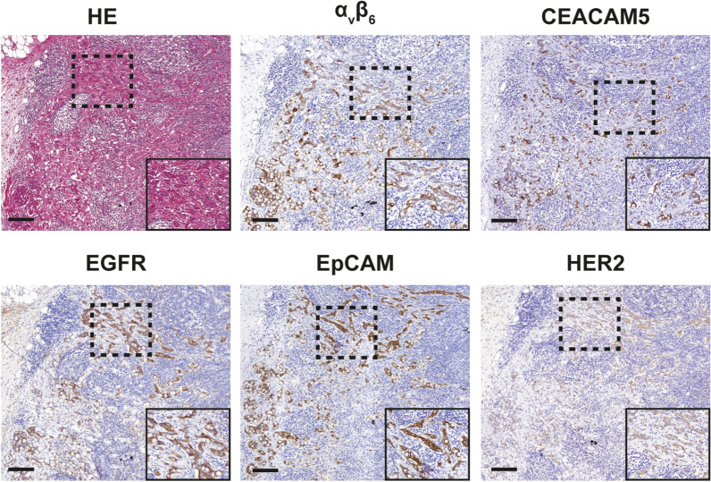
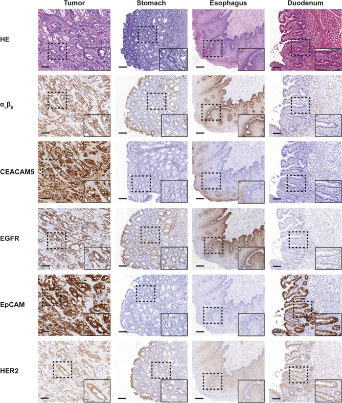
Results: Positive biomarker expression in primary gastric tumors was observed in 86% for αvβ6, 72% for CEACAM5, 77% for EGFR, 93% for EpCAM and 71% for HER2. Tumor expression of CEACAM5, EGFR and EpCAM was higher compared to healthy stomach tissue expression, while this was not the case for αvβ6 and HER2. Tumor-positive lymph nodes could be distinguished from tumor-negative lymph nodes, with accuracy ranging from 82 to 93% between biomarkers. CEACAM5, EGFR and EpCAM were abundantly expressed on distant metastases, with expression in 88-95% of tissue specimens.
Conclusion: Our findings show that CEACAM5, EGFR and EpCAM are promising targets for molecular imaging of primary gastric cancer, as well as visualization of both lymph node and distant metastases. Further clinical evaluation of PET and FGS tracers targeting these antigens is warranted.
期刊介绍:
Molecular Diagnosis & Therapy welcomes current opinion articles on emerging or contentious issues, comprehensive narrative reviews, systematic reviews (as outlined by the PRISMA statement), original research articles (including short communications) and letters to the editor. All manuscripts are subject to peer review by international experts.
 求助内容:
求助内容: 应助结果提醒方式:


