Zheyuan Zhang , Elif Keles , Gorkem Durak , Yavuz Taktak , Onkar Susladkar , Vandan Gorade , Debesh Jha , Asli C. Ormeci , Alpay Medetalibeyoglu , Lanhong Yao , Bin Wang , Ilkin Sevgi Isler , Linkai Peng , Hongyi Pan , Camila Lopes Vendrami , Amir Bourhani , Yury Velichko , Boqing Gong , Concetto Spampinato , Ayis Pyrros , Ulas Bagci
{"title":"利用深度学习对胰腺进行大规模多中心 CT 和 MRI 分割。","authors":"Zheyuan Zhang , Elif Keles , Gorkem Durak , Yavuz Taktak , Onkar Susladkar , Vandan Gorade , Debesh Jha , Asli C. Ormeci , Alpay Medetalibeyoglu , Lanhong Yao , Bin Wang , Ilkin Sevgi Isler , Linkai Peng , Hongyi Pan , Camila Lopes Vendrami , Amir Bourhani , Yury Velichko , Boqing Gong , Concetto Spampinato , Ayis Pyrros , Ulas Bagci","doi":"10.1016/j.media.2024.103382","DOIUrl":null,"url":null,"abstract":"<div><div>Automated volumetric segmentation of the pancreas on cross-sectional imaging is needed for diagnosis and follow-up of pancreatic diseases. While CT-based pancreatic segmentation is more established, MRI-based segmentation methods are understudied, largely due to a lack of publicly available datasets, benchmarking research efforts, and domain-specific deep learning methods. In this retrospective study, we collected a large dataset (767 scans from 499 participants) of T1-weighted (T1 W) and T2-weighted (T2 W) abdominal MRI series from five centers between March 2004 and November 2022. We also collected CT scans of 1,350 patients from publicly available sources for benchmarking purposes. We introduced a new pancreas segmentation method, called <em>PanSegNet</em>, combining the strengths of <em>nnUNet</em> and a <em>Transformer</em> network with a new linear attention module enabling volumetric computation. We tested <em>PanSegNet</em>’s accuracy in cross-modality (a total of 2,117 scans) and cross-center settings with Dice and Hausdorff distance (HD95) evaluation metrics. We used Cohen’s kappa statistics for intra and inter-rater agreement evaluation and paired t-tests for volume and Dice comparisons, respectively. For segmentation accuracy, we achieved Dice coefficients of 88.3% (±7.2%, at case level) with CT, 85.0% (±7.9%) with T1 W MRI, and 86.3% (±6.4%) with T2 W MRI. There was a high correlation for pancreas volume prediction with <span><math><msup><mrow><mi>R</mi></mrow><mrow><mn>2</mn></mrow></msup></math></span> of 0.91, 0.84, and 0.85 for CT, T1 W, and T2 W, respectively. We found moderate inter-observer (0.624 and 0.638 for T1 W and T2 W MRI, respectively) and high intra-observer agreement scores. All MRI data is made available at <span><span>https://osf.io/kysnj/</span><svg><path></path></svg></span>. Our source code is available at <span><span>https://github.com/NUBagciLab/PaNSegNet</span><svg><path></path></svg></span>.</div></div>","PeriodicalId":18328,"journal":{"name":"Medical image analysis","volume":"99 ","pages":"Article 103382"},"PeriodicalIF":10.7000,"publicationDate":"2024-11-08","publicationTypes":"Journal Article","fieldsOfStudy":null,"isOpenAccess":false,"openAccessPdf":"","citationCount":"0","resultStr":"{\"title\":\"Large-scale multi-center CT and MRI segmentation of pancreas with deep learning\",\"authors\":\"Zheyuan Zhang , Elif Keles , Gorkem Durak , Yavuz Taktak , Onkar Susladkar , Vandan Gorade , Debesh Jha , Asli C. Ormeci , Alpay Medetalibeyoglu , Lanhong Yao , Bin Wang , Ilkin Sevgi Isler , Linkai Peng , Hongyi Pan , Camila Lopes Vendrami , Amir Bourhani , Yury Velichko , Boqing Gong , Concetto Spampinato , Ayis Pyrros , Ulas Bagci\",\"doi\":\"10.1016/j.media.2024.103382\",\"DOIUrl\":null,\"url\":null,\"abstract\":\"<div><div>Automated volumetric segmentation of the pancreas on cross-sectional imaging is needed for diagnosis and follow-up of pancreatic diseases. While CT-based pancreatic segmentation is more established, MRI-based segmentation methods are understudied, largely due to a lack of publicly available datasets, benchmarking research efforts, and domain-specific deep learning methods. In this retrospective study, we collected a large dataset (767 scans from 499 participants) of T1-weighted (T1 W) and T2-weighted (T2 W) abdominal MRI series from five centers between March 2004 and November 2022. We also collected CT scans of 1,350 patients from publicly available sources for benchmarking purposes. We introduced a new pancreas segmentation method, called <em>PanSegNet</em>, combining the strengths of <em>nnUNet</em> and a <em>Transformer</em> network with a new linear attention module enabling volumetric computation. We tested <em>PanSegNet</em>’s accuracy in cross-modality (a total of 2,117 scans) and cross-center settings with Dice and Hausdorff distance (HD95) evaluation metrics. We used Cohen’s kappa statistics for intra and inter-rater agreement evaluation and paired t-tests for volume and Dice comparisons, respectively. For segmentation accuracy, we achieved Dice coefficients of 88.3% (±7.2%, at case level) with CT, 85.0% (±7.9%) with T1 W MRI, and 86.3% (±6.4%) with T2 W MRI. There was a high correlation for pancreas volume prediction with <span><math><msup><mrow><mi>R</mi></mrow><mrow><mn>2</mn></mrow></msup></math></span> of 0.91, 0.84, and 0.85 for CT, T1 W, and T2 W, respectively. We found moderate inter-observer (0.624 and 0.638 for T1 W and T2 W MRI, respectively) and high intra-observer agreement scores. All MRI data is made available at <span><span>https://osf.io/kysnj/</span><svg><path></path></svg></span>. Our source code is available at <span><span>https://github.com/NUBagciLab/PaNSegNet</span><svg><path></path></svg></span>.</div></div>\",\"PeriodicalId\":18328,\"journal\":{\"name\":\"Medical image analysis\",\"volume\":\"99 \",\"pages\":\"Article 103382\"},\"PeriodicalIF\":10.7000,\"publicationDate\":\"2024-11-08\",\"publicationTypes\":\"Journal Article\",\"fieldsOfStudy\":null,\"isOpenAccess\":false,\"openAccessPdf\":\"\",\"citationCount\":\"0\",\"resultStr\":null,\"platform\":\"Semanticscholar\",\"paperid\":null,\"PeriodicalName\":\"Medical image analysis\",\"FirstCategoryId\":\"5\",\"ListUrlMain\":\"https://www.sciencedirect.com/science/article/pii/S1361841524003074\",\"RegionNum\":1,\"RegionCategory\":\"医学\",\"ArticlePicture\":[],\"TitleCN\":null,\"AbstractTextCN\":null,\"PMCID\":null,\"EPubDate\":\"\",\"PubModel\":\"\",\"JCR\":\"Q1\",\"JCRName\":\"COMPUTER SCIENCE, ARTIFICIAL INTELLIGENCE\",\"Score\":null,\"Total\":0}","platform":"Semanticscholar","paperid":null,"PeriodicalName":"Medical image analysis","FirstCategoryId":"5","ListUrlMain":"https://www.sciencedirect.com/science/article/pii/S1361841524003074","RegionNum":1,"RegionCategory":"医学","ArticlePicture":[],"TitleCN":null,"AbstractTextCN":null,"PMCID":null,"EPubDate":"","PubModel":"","JCR":"Q1","JCRName":"COMPUTER SCIENCE, ARTIFICIAL INTELLIGENCE","Score":null,"Total":0}
Large-scale multi-center CT and MRI segmentation of pancreas with deep learning
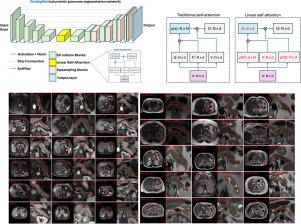
Automated volumetric segmentation of the pancreas on cross-sectional imaging is needed for diagnosis and follow-up of pancreatic diseases. While CT-based pancreatic segmentation is more established, MRI-based segmentation methods are understudied, largely due to a lack of publicly available datasets, benchmarking research efforts, and domain-specific deep learning methods. In this retrospective study, we collected a large dataset (767 scans from 499 participants) of T1-weighted (T1 W) and T2-weighted (T2 W) abdominal MRI series from five centers between March 2004 and November 2022. We also collected CT scans of 1,350 patients from publicly available sources for benchmarking purposes. We introduced a new pancreas segmentation method, called PanSegNet, combining the strengths of nnUNet and a Transformer network with a new linear attention module enabling volumetric computation. We tested PanSegNet’s accuracy in cross-modality (a total of 2,117 scans) and cross-center settings with Dice and Hausdorff distance (HD95) evaluation metrics. We used Cohen’s kappa statistics for intra and inter-rater agreement evaluation and paired t-tests for volume and Dice comparisons, respectively. For segmentation accuracy, we achieved Dice coefficients of 88.3% (±7.2%, at case level) with CT, 85.0% (±7.9%) with T1 W MRI, and 86.3% (±6.4%) with T2 W MRI. There was a high correlation for pancreas volume prediction with of 0.91, 0.84, and 0.85 for CT, T1 W, and T2 W, respectively. We found moderate inter-observer (0.624 and 0.638 for T1 W and T2 W MRI, respectively) and high intra-observer agreement scores. All MRI data is made available at https://osf.io/kysnj/. Our source code is available at https://github.com/NUBagciLab/PaNSegNet.
期刊介绍:
Medical Image Analysis serves as a platform for sharing new research findings in the realm of medical and biological image analysis, with a focus on applications of computer vision, virtual reality, and robotics to biomedical imaging challenges. The journal prioritizes the publication of high-quality, original papers contributing to the fundamental science of processing, analyzing, and utilizing medical and biological images. It welcomes approaches utilizing biomedical image datasets across all spatial scales, from molecular/cellular imaging to tissue/organ imaging.