CSGO:用于血沉和伊红染色组织全细胞分割的深度学习管道。

IF 5.1 2区 医学 Q1 MEDICINE, RESEARCH & EXPERIMENTAL
Zifan Gu, Shidan Wang, Ruichen Rong, Zhuo Zhao, Fangjiang Wu, Qin Zhou, Zhuoyu Wen, Zhikai Chi, Yisheng Fang, Yan Peng, Liwei Jia, Mingyi Chen, Donghan M Yang, Yujin Hoshida, Yang Xie, Guanghua Xiao
{"title":"CSGO:用于血沉和伊红染色组织全细胞分割的深度学习管道。","authors":"Zifan Gu, Shidan Wang, Ruichen Rong, Zhuo Zhao, Fangjiang Wu, Qin Zhou, Zhuoyu Wen, Zhikai Chi, Yisheng Fang, Yan Peng, Liwei Jia, Mingyi Chen, Donghan M Yang, Yujin Hoshida, Yang Xie, Guanghua Xiao","doi":"10.1016/j.labinv.2024.102184","DOIUrl":null,"url":null,"abstract":"<p><p>Accurate whole-cell segmentation is essential in various biomedical applications, particularly in studying the tumor microenvironment (TME). Despite advancements in machine learning for nuclei segmentation in hematoxylin and eosin (H&E) stained images, there remains a need for effective whole-cell segmentation methods. This study aims to develop a deep learning-based pipeline to automatically segment cells in H&E-stained tissues, thereby advancing the capabilities of pathological image analysis. The Cell Segmentation with Globally Optimized boundaries (CSGO) framework integrates nuclei and membrane segmentation algorithms, followed by post-processing using an energy-based watershed method. Specifically, we employed the You Only Look Once (Yolo) object detection algorithm for nuclei segmentation and U-Net for membrane segmentation. The membrane detection model was trained on a dataset of 7 hepatocellular carcinomas and 11 normal liver tissue patches. The cell segmentation performance was extensively evaluated on five external datasets, including liver, lung, and oral disease cases. CSGO demonstrated superior performance over the state-of-the-art method Cellpose, achieving higher F1 scores ranging from 0.37 to 0.53 at an intersection over union (IoU) threshold of 0.5 in four of the five external datasets, compared to that of Cellpose from 0.21 to 0.36. These results underscore the robustness and accuracy of our approach in various tissue types. A web-based application is available at https://ai.swmed.edu/projects/csgo, providing a user-friendly platform for researchers to apply our method to their own datasets. Our method exhibits remarkable versatility in whole-cell segmentation across diverse cancer subtypes, serving as an accurate and reliable tool to facilitate TME studies. The advancements presented in this study have the potential to significantly enhance the precision and efficiency of pathological image analysis, contributing to better understanding and treatment of cancer.</p>","PeriodicalId":17930,"journal":{"name":"Laboratory Investigation","volume":" ","pages":"102184"},"PeriodicalIF":5.1000,"publicationDate":"2024-11-09","publicationTypes":"Journal Article","fieldsOfStudy":null,"isOpenAccess":false,"openAccessPdf":"","citationCount":"0","resultStr":"{\"title\":\"CSGO: A Deep Learning Pipeline for Whole-Cell Segmentation in Hematoxylin and Eosin Stained Tissues.\",\"authors\":\"Zifan Gu, Shidan Wang, Ruichen Rong, Zhuo Zhao, Fangjiang Wu, Qin Zhou, Zhuoyu Wen, Zhikai Chi, Yisheng Fang, Yan Peng, Liwei Jia, Mingyi Chen, Donghan M Yang, Yujin Hoshida, Yang Xie, Guanghua Xiao\",\"doi\":\"10.1016/j.labinv.2024.102184\",\"DOIUrl\":null,\"url\":null,\"abstract\":\"<p><p>Accurate whole-cell segmentation is essential in various biomedical applications, particularly in studying the tumor microenvironment (TME). Despite advancements in machine learning for nuclei segmentation in hematoxylin and eosin (H&E) stained images, there remains a need for effective whole-cell segmentation methods. This study aims to develop a deep learning-based pipeline to automatically segment cells in H&E-stained tissues, thereby advancing the capabilities of pathological image analysis. The Cell Segmentation with Globally Optimized boundaries (CSGO) framework integrates nuclei and membrane segmentation algorithms, followed by post-processing using an energy-based watershed method. Specifically, we employed the You Only Look Once (Yolo) object detection algorithm for nuclei segmentation and U-Net for membrane segmentation. The membrane detection model was trained on a dataset of 7 hepatocellular carcinomas and 11 normal liver tissue patches. The cell segmentation performance was extensively evaluated on five external datasets, including liver, lung, and oral disease cases. CSGO demonstrated superior performance over the state-of-the-art method Cellpose, achieving higher F1 scores ranging from 0.37 to 0.53 at an intersection over union (IoU) threshold of 0.5 in four of the five external datasets, compared to that of Cellpose from 0.21 to 0.36. These results underscore the robustness and accuracy of our approach in various tissue types. A web-based application is available at https://ai.swmed.edu/projects/csgo, providing a user-friendly platform for researchers to apply our method to their own datasets. Our method exhibits remarkable versatility in whole-cell segmentation across diverse cancer subtypes, serving as an accurate and reliable tool to facilitate TME studies. The advancements presented in this study have the potential to significantly enhance the precision and efficiency of pathological image analysis, contributing to better understanding and treatment of cancer.</p>\",\"PeriodicalId\":17930,\"journal\":{\"name\":\"Laboratory Investigation\",\"volume\":\" \",\"pages\":\"102184\"},\"PeriodicalIF\":5.1000,\"publicationDate\":\"2024-11-09\",\"publicationTypes\":\"Journal Article\",\"fieldsOfStudy\":null,\"isOpenAccess\":false,\"openAccessPdf\":\"\",\"citationCount\":\"0\",\"resultStr\":null,\"platform\":\"Semanticscholar\",\"paperid\":null,\"PeriodicalName\":\"Laboratory Investigation\",\"FirstCategoryId\":\"3\",\"ListUrlMain\":\"https://doi.org/10.1016/j.labinv.2024.102184\",\"RegionNum\":2,\"RegionCategory\":\"医学\",\"ArticlePicture\":[],\"TitleCN\":null,\"AbstractTextCN\":null,\"PMCID\":null,\"EPubDate\":\"\",\"PubModel\":\"\",\"JCR\":\"Q1\",\"JCRName\":\"MEDICINE, RESEARCH & EXPERIMENTAL\",\"Score\":null,\"Total\":0}","platform":"Semanticscholar","paperid":null,"PeriodicalName":"Laboratory Investigation","FirstCategoryId":"3","ListUrlMain":"https://doi.org/10.1016/j.labinv.2024.102184","RegionNum":2,"RegionCategory":"医学","ArticlePicture":[],"TitleCN":null,"AbstractTextCN":null,"PMCID":null,"EPubDate":"","PubModel":"","JCR":"Q1","JCRName":"MEDICINE, RESEARCH & EXPERIMENTAL","Score":null,"Total":0}
引用次数: 0

摘要

在各种生物医学应用中,尤其是在研究肿瘤微环境(TME)时,准确的全细胞分割至关重要。尽管机器学习在苏木精和伊红(H&E)染色图像的细胞核分割方面取得了进步,但仍然需要有效的全细胞分割方法。本研究旨在开发一种基于深度学习的管道,自动分割 H&E 染色组织中的细胞,从而提高病理图像分析能力。细胞全局优化边界分割(CSGO)框架集成了细胞核和细胞膜分割算法,然后使用基于能量的分水岭方法进行后处理。具体来说,我们采用 "你只看一次(Yolo)"对象检测算法进行细胞核分割,采用 U-Net 进行细胞膜分割。膜检测模型是在 7 个肝细胞癌和 11 个正常肝组织斑块的数据集上训练出来的。在五个外部数据集(包括肝脏、肺部和口腔疾病病例)上对细胞分割性能进行了广泛评估。与最先进的 Cellpose 方法相比,CSGO 表现出更优越的性能,在五个外部数据集中的四个数据集中,当交集大于联合(IoU)阈值为 0.5 时,CSGO 获得了 0.37 至 0.53 的更高 F1 分数,而 Cellpose 的分数为 0.21 至 0.36。这些结果凸显了我们的方法在各种组织类型中的稳健性和准确性。https://ai.swmed.edu/projects/csgo 网站提供了一个基于网络的应用程序,为研究人员将我们的方法应用于自己的数据集提供了一个用户友好型平台。我们的方法在不同癌症亚型的全细胞分割中表现出卓越的多功能性,是促进 TME 研究的准确可靠的工具。本研究中介绍的进展有可能大大提高病理图像分析的精确度和效率,有助于更好地理解和治疗癌症。
本文章由计算机程序翻译,如有差异,请以英文原文为准。
CSGO: A Deep Learning Pipeline for Whole-Cell Segmentation in Hematoxylin and Eosin Stained Tissues.

Accurate whole-cell segmentation is essential in various biomedical applications, particularly in studying the tumor microenvironment (TME). Despite advancements in machine learning for nuclei segmentation in hematoxylin and eosin (H&E) stained images, there remains a need for effective whole-cell segmentation methods. This study aims to develop a deep learning-based pipeline to automatically segment cells in H&E-stained tissues, thereby advancing the capabilities of pathological image analysis. The Cell Segmentation with Globally Optimized boundaries (CSGO) framework integrates nuclei and membrane segmentation algorithms, followed by post-processing using an energy-based watershed method. Specifically, we employed the You Only Look Once (Yolo) object detection algorithm for nuclei segmentation and U-Net for membrane segmentation. The membrane detection model was trained on a dataset of 7 hepatocellular carcinomas and 11 normal liver tissue patches. The cell segmentation performance was extensively evaluated on five external datasets, including liver, lung, and oral disease cases. CSGO demonstrated superior performance over the state-of-the-art method Cellpose, achieving higher F1 scores ranging from 0.37 to 0.53 at an intersection over union (IoU) threshold of 0.5 in four of the five external datasets, compared to that of Cellpose from 0.21 to 0.36. These results underscore the robustness and accuracy of our approach in various tissue types. A web-based application is available at https://ai.swmed.edu/projects/csgo, providing a user-friendly platform for researchers to apply our method to their own datasets. Our method exhibits remarkable versatility in whole-cell segmentation across diverse cancer subtypes, serving as an accurate and reliable tool to facilitate TME studies. The advancements presented in this study have the potential to significantly enhance the precision and efficiency of pathological image analysis, contributing to better understanding and treatment of cancer.

求助全文
通过发布文献求助,成功后即可免费获取论文全文。 去求助
来源期刊
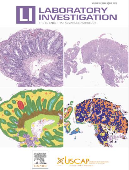
Laboratory Investigation
Laboratory Investigation 医学-病理学
CiteScore
8.30
自引率
0.00%
发文量
125
审稿时长
2 months
期刊介绍: Laboratory Investigation is an international journal owned by the United States and Canadian Academy of Pathology. Laboratory Investigation offers prompt publication of high-quality original research in all biomedical disciplines relating to the understanding of human disease and the application of new methods to the diagnosis of disease. Both human and experimental studies are welcome.
×
引用
GB/T 7714-2015
复制
MLA
复制
APA
复制
导出至
BibTeX EndNote RefMan NoteFirst NoteExpress
×
提示
您的信息不完整,为了账户安全,请先补充。
现在去补充
×
提示
您因"违规操作"
具体请查看互助需知
我知道了
×
提示
确定
请完成安全验证×
copy
已复制链接
快去分享给好友吧!
我知道了
右上角分享
点击右上角分享
0
联系我们:info@booksci.cn Book学术提供免费学术资源搜索服务,方便国内外学者检索中英文文献。致力于提供最便捷和优质的服务体验。 Copyright © 2023 布克学术 All rights reserved.
京ICP备2023020795号-1
ghs 京公网安备 11010802042870号
Book学术文献互助
Book学术文献互助群
群 号:481959085
Book学术官方微信