蛋白 tau 在富脯氨酸区的磷酸化及其对阿尔茨海默氏症进展的影响。

IF 4.6 2区 医学 Q1 NEUROSCIENCES
Paula Merino-Serrais , José Miguel Soria , Cristina Aguirre Arrabal , Alfonso Ortigado-López , María Ángeles García Esparza , Alberto Muñoz , Félix Hernández , Jesús Ávila , Javier DeFelipe , Gonzalo León-Espinosa
{"title":"蛋白 tau 在富脯氨酸区的磷酸化及其对阿尔茨海默氏症进展的影响。","authors":"Paula Merino-Serrais ,&nbsp;José Miguel Soria ,&nbsp;Cristina Aguirre Arrabal ,&nbsp;Alfonso Ortigado-López ,&nbsp;María Ángeles García Esparza ,&nbsp;Alberto Muñoz ,&nbsp;Félix Hernández ,&nbsp;Jesús Ávila ,&nbsp;Javier DeFelipe ,&nbsp;Gonzalo León-Espinosa","doi":"10.1016/j.expneurol.2024.115049","DOIUrl":null,"url":null,"abstract":"<div><div>Tau has a wide variety of essential functions in the brain, but this protein also plays a determining role in the development of Alzheimer's disease (AD) and other neurodegenerative diseases called tauopathies. This is due to its abnormal aggregation and the subsequent formation of neurofibrillary tangles. Tau hyperphosphorylation appears to be a critical step in its transformation into an aggregated protein. However, the exact process, including the cellular events that trigger it, remains unclear. In this study, we employed immunocytochemistry assays on hippocampal sections from AD cases and from tauopathy cases (Braak stage III) with no evidence of cognitive decline, and the P301S mouse model to investigate the colocalization patterns of Tau phosphorylated (p) at specific residues (S202-T205, S214, and T231) within the proline-rich region. Our results show pyramidal neurons in the hippocampus of P301S mice in which Tau is intensely phosphorylated at residues S202 and T205 (recognized by the AT8 antibody), but with no detectable phosphorylation at S214 or T231. These non-colocalizing neurons displayed intensely labeled aggregated pTau deposits distributed through the soma and dendritic processes. However, most of the hippocampal pyramidal neurons are labeled with pTauS214 or pTauT231 antibodies and typically showed a homogeneous and diffuse pTau distribution (not aggregated). This different labeling likely reflects a Tau conformational step, potentially related to the transition from a diffuse tau phosphorylation phenotype (Type 2) into an NFT-like or Type 1 phenotype. We further observed that dendrites of CA3 pyramidal cells are intensely labeled with pTau214 in the <em>stratum lucidum</em>, but not with AT8 or pTauT231. By contrast, analysis of tissue from AD patients or other human tauopathy cases (Braak stage III) with no evidence of cognitive decline revealed extensive colocalization with both antibody combinations in CA1. The complete or mature tangle development may follow a different mechanism in the P301S mouse model or may require more time to achieve the maturity state found in AD cases. Further studies would be necessary to address this question.</div></div>","PeriodicalId":12246,"journal":{"name":"Experimental Neurology","volume":"383 ","pages":"Article 115049"},"PeriodicalIF":4.6000,"publicationDate":"2024-11-08","publicationTypes":"Journal Article","fieldsOfStudy":null,"isOpenAccess":false,"openAccessPdf":"","citationCount":"0","resultStr":"{\"title\":\"Protein tau phosphorylation in the proline rich region and its implication in the progression of Alzheimer's disease\",\"authors\":\"Paula Merino-Serrais ,&nbsp;José Miguel Soria ,&nbsp;Cristina Aguirre Arrabal ,&nbsp;Alfonso Ortigado-López ,&nbsp;María Ángeles García Esparza ,&nbsp;Alberto Muñoz ,&nbsp;Félix Hernández ,&nbsp;Jesús Ávila ,&nbsp;Javier DeFelipe ,&nbsp;Gonzalo León-Espinosa\",\"doi\":\"10.1016/j.expneurol.2024.115049\",\"DOIUrl\":null,\"url\":null,\"abstract\":\"<div><div>Tau has a wide variety of essential functions in the brain, but this protein also plays a determining role in the development of Alzheimer's disease (AD) and other neurodegenerative diseases called tauopathies. This is due to its abnormal aggregation and the subsequent formation of neurofibrillary tangles. Tau hyperphosphorylation appears to be a critical step in its transformation into an aggregated protein. However, the exact process, including the cellular events that trigger it, remains unclear. In this study, we employed immunocytochemistry assays on hippocampal sections from AD cases and from tauopathy cases (Braak stage III) with no evidence of cognitive decline, and the P301S mouse model to investigate the colocalization patterns of Tau phosphorylated (p) at specific residues (S202-T205, S214, and T231) within the proline-rich region. Our results show pyramidal neurons in the hippocampus of P301S mice in which Tau is intensely phosphorylated at residues S202 and T205 (recognized by the AT8 antibody), but with no detectable phosphorylation at S214 or T231. These non-colocalizing neurons displayed intensely labeled aggregated pTau deposits distributed through the soma and dendritic processes. However, most of the hippocampal pyramidal neurons are labeled with pTauS214 or pTauT231 antibodies and typically showed a homogeneous and diffuse pTau distribution (not aggregated). This different labeling likely reflects a Tau conformational step, potentially related to the transition from a diffuse tau phosphorylation phenotype (Type 2) into an NFT-like or Type 1 phenotype. We further observed that dendrites of CA3 pyramidal cells are intensely labeled with pTau214 in the <em>stratum lucidum</em>, but not with AT8 or pTauT231. By contrast, analysis of tissue from AD patients or other human tauopathy cases (Braak stage III) with no evidence of cognitive decline revealed extensive colocalization with both antibody combinations in CA1. The complete or mature tangle development may follow a different mechanism in the P301S mouse model or may require more time to achieve the maturity state found in AD cases. Further studies would be necessary to address this question.</div></div>\",\"PeriodicalId\":12246,\"journal\":{\"name\":\"Experimental Neurology\",\"volume\":\"383 \",\"pages\":\"Article 115049\"},\"PeriodicalIF\":4.6000,\"publicationDate\":\"2024-11-08\",\"publicationTypes\":\"Journal Article\",\"fieldsOfStudy\":null,\"isOpenAccess\":false,\"openAccessPdf\":\"\",\"citationCount\":\"0\",\"resultStr\":null,\"platform\":\"Semanticscholar\",\"paperid\":null,\"PeriodicalName\":\"Experimental Neurology\",\"FirstCategoryId\":\"3\",\"ListUrlMain\":\"https://www.sciencedirect.com/science/article/pii/S0014488624003753\",\"RegionNum\":2,\"RegionCategory\":\"医学\",\"ArticlePicture\":[],\"TitleCN\":null,\"AbstractTextCN\":null,\"PMCID\":null,\"EPubDate\":\"\",\"PubModel\":\"\",\"JCR\":\"Q1\",\"JCRName\":\"NEUROSCIENCES\",\"Score\":null,\"Total\":0}","platform":"Semanticscholar","paperid":null,"PeriodicalName":"Experimental Neurology","FirstCategoryId":"3","ListUrlMain":"https://www.sciencedirect.com/science/article/pii/S0014488624003753","RegionNum":2,"RegionCategory":"医学","ArticlePicture":[],"TitleCN":null,"AbstractTextCN":null,"PMCID":null,"EPubDate":"","PubModel":"","JCR":"Q1","JCRName":"NEUROSCIENCES","Score":null,"Total":0}
引用次数: 0

摘要

Tau 蛋白在大脑中具有多种重要功能,但这种蛋白质在阿尔茨海默病(AD)和其他称为 Tau 病的神经退行性疾病的发病过程中也起着决定性作用。这是由于它的异常聚集和随后形成的神经纤维缠结。Tau 过度磷酸化似乎是其转变为聚集蛋白的关键步骤。然而,包括引发该过程的细胞事件在内的确切过程仍不清楚。在本研究中,我们采用免疫细胞化学法检测了来自AD病例和无认知功能衰退迹象的tauopathy病例(Braak III期)以及P301S小鼠模型的海马切片,研究了Tau在富脯氨酸区域内特定残基(S202-T205、S214和T231)磷酸化(p)的共定位模式。我们的结果显示,在 P301S 小鼠海马的锥体神经元中,Tau 在残基 S202 和 T205(AT8 抗体可识别)处发生了强烈的磷酸化,但在 S214 或 T231 处却检测不到磷酸化。这些非定位神经元显示出强烈标记的聚合 pTau 沉积,分布在体节和树突过程中。然而,大多数海马锥体神经元都用 pTauS214 或 pTauT231 抗体进行了标记,通常显示出均匀和弥散的 pTau 分布(非聚集)。这种不同的标记可能反映了 Tau 的构象步骤,可能与从弥散的 tau 磷酸化表型(2 型)过渡到类似 NFT 或 1 型表型有关。我们进一步观察到,CA3锥体细胞的树突在透明层中被pTau214强烈标记,但没有被AT8或pTauT231标记。相比之下,对无认知能力下降迹象的AD患者或其他人类tauopathy病例(Braak III期)的组织进行分析后发现,这两种抗体组合在CA1有广泛的共定位。在 P301S 小鼠模型中,完全或成熟的纠结发育可能遵循不同的机制,或者需要更多的时间才能达到 AD 病例中的成熟状态。要解决这个问题,还需要进一步的研究。
本文章由计算机程序翻译,如有差异,请以英文原文为准。
Protein tau phosphorylation in the proline rich region and its implication in the progression of Alzheimer's disease
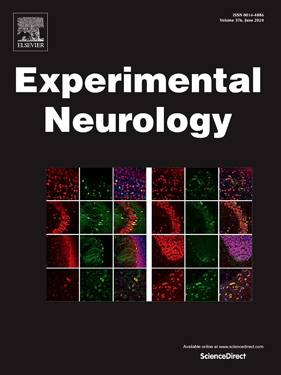
Tau has a wide variety of essential functions in the brain, but this protein also plays a determining role in the development of Alzheimer's disease (AD) and other neurodegenerative diseases called tauopathies. This is due to its abnormal aggregation and the subsequent formation of neurofibrillary tangles. Tau hyperphosphorylation appears to be a critical step in its transformation into an aggregated protein. However, the exact process, including the cellular events that trigger it, remains unclear. In this study, we employed immunocytochemistry assays on hippocampal sections from AD cases and from tauopathy cases (Braak stage III) with no evidence of cognitive decline, and the P301S mouse model to investigate the colocalization patterns of Tau phosphorylated (p) at specific residues (S202-T205, S214, and T231) within the proline-rich region. Our results show pyramidal neurons in the hippocampus of P301S mice in which Tau is intensely phosphorylated at residues S202 and T205 (recognized by the AT8 antibody), but with no detectable phosphorylation at S214 or T231. These non-colocalizing neurons displayed intensely labeled aggregated pTau deposits distributed through the soma and dendritic processes. However, most of the hippocampal pyramidal neurons are labeled with pTauS214 or pTauT231 antibodies and typically showed a homogeneous and diffuse pTau distribution (not aggregated). This different labeling likely reflects a Tau conformational step, potentially related to the transition from a diffuse tau phosphorylation phenotype (Type 2) into an NFT-like or Type 1 phenotype. We further observed that dendrites of CA3 pyramidal cells are intensely labeled with pTau214 in the stratum lucidum, but not with AT8 or pTauT231. By contrast, analysis of tissue from AD patients or other human tauopathy cases (Braak stage III) with no evidence of cognitive decline revealed extensive colocalization with both antibody combinations in CA1. The complete or mature tangle development may follow a different mechanism in the P301S mouse model or may require more time to achieve the maturity state found in AD cases. Further studies would be necessary to address this question.
求助全文
通过发布文献求助,成功后即可免费获取论文全文。 去求助
来源期刊
Experimental Neurology
Experimental Neurology 医学-神经科学
CiteScore
10.10
自引率
3.80%
发文量
258
审稿时长
42 days
期刊介绍: Experimental Neurology, a Journal of Neuroscience Research, publishes original research in neuroscience with a particular emphasis on novel findings in neural development, regeneration, plasticity and transplantation. The journal has focused on research concerning basic mechanisms underlying neurological disorders.
×
引用
GB/T 7714-2015
复制
MLA
复制
APA
复制
导出至
BibTeX EndNote RefMan NoteFirst NoteExpress
×
提示
您的信息不完整,为了账户安全,请先补充。
现在去补充
×
提示
您因"违规操作"
具体请查看互助需知
我知道了
×
提示
确定
请完成安全验证×
copy
已复制链接
快去分享给好友吧!
我知道了
右上角分享
点击右上角分享
0
联系我们:info@booksci.cn Book学术提供免费学术资源搜索服务,方便国内外学者检索中英文文献。致力于提供最便捷和优质的服务体验。 Copyright © 2023 布克学术 All rights reserved.
京ICP备2023020795号-1
ghs 京公网安备 11010802042870号
Book学术文献互助
Book学术文献互助群
群 号:481959085
Book学术官方微信