Denis Cyr, Michel Boutin, Bruno Maranda, Paula J. Waters
{"title":"加强对 3-羟基戊二酸和 2-羟基戊二酸的区分有助于对戊二酸尿症 1 型进行诊断检测。","authors":"Denis Cyr, Michel Boutin, Bruno Maranda, Paula J. Waters","doi":"10.1002/jmd2.12447","DOIUrl":null,"url":null,"abstract":"<p>Glutaric aciduria type 1 (GA1) is an inherited neurometabolic disorder, in which deficiency of glutaryl-CoA dehydrogenase leads to accumulation of glutaric acid (GA) and 3-hydroxyglutaric acid (3-HG). Some low excretors may exhibit only slight elevation of urinary 3-HG, with normal urinary GA, yet are at significant risk of severe clinical disease. Accurate quantitation of urinary 3-HG is crucial in diagnostic workup for GA1, but in this context, current gas chromatography–mass spectrometry (GC–MS) methods have inherent analytical challenges. Co-elution and spectral similarities of the 3-HG and 2-HG structural isomers can cause difficulties in quantitation of slightly elevated 3-HG. Our laboratory recently acquired a gas chromatography system coupled to a triple quadrupole mass spectrometer (GC–MS/MS), and we took advantage of its increased sensitivity and specificity to improve our existing GC–MS method. A stable isotope dilution process is used, with sample treatment consisting of a double liquid–liquid extraction followed by a trimethylsilyl derivatization. The transitions <i>m</i>/<i>z</i> 349 → 333 for 3-HG and <i>m</i>/<i>z</i> 349 → 321 for 2-HG were selected to differentiate these two isobaric molecules based on their characteristic fragments, thus minimizing interferences despite co-elution. Method validation demonstrated satisfactory precision and accuracy. Using GC–MS/MS instead of GC–MS allowed us to decrease the required specimen volume, number of sample processing steps, chromatographic run time, and instrument maintenance. This enhanced assay facilitates clinical laboratory testing for GA1, both in confirmatory protocols following positive newborn screening and in diagnostic investigation of patients with suggestive signs or symptoms.</p>","PeriodicalId":14930,"journal":{"name":"JIMD reports","volume":"65 6","pages":"433-441"},"PeriodicalIF":1.8000,"publicationDate":"2024-08-27","publicationTypes":"Journal Article","fieldsOfStudy":null,"isOpenAccess":false,"openAccessPdf":"https://www.ncbi.nlm.nih.gov/pmc/articles/PMC11540565/pdf/","citationCount":"0","resultStr":"{\"title\":\"Enhanced differentiation between 3-hydroxyglutaric and 2-hydroxyglutaric acids facilitates diagnostic testing for glutaric aciduria type 1\",\"authors\":\"Denis Cyr, Michel Boutin, Bruno Maranda, Paula J. Waters\",\"doi\":\"10.1002/jmd2.12447\",\"DOIUrl\":null,\"url\":null,\"abstract\":\"<p>Glutaric aciduria type 1 (GA1) is an inherited neurometabolic disorder, in which deficiency of glutaryl-CoA dehydrogenase leads to accumulation of glutaric acid (GA) and 3-hydroxyglutaric acid (3-HG). Some low excretors may exhibit only slight elevation of urinary 3-HG, with normal urinary GA, yet are at significant risk of severe clinical disease. Accurate quantitation of urinary 3-HG is crucial in diagnostic workup for GA1, but in this context, current gas chromatography–mass spectrometry (GC–MS) methods have inherent analytical challenges. Co-elution and spectral similarities of the 3-HG and 2-HG structural isomers can cause difficulties in quantitation of slightly elevated 3-HG. Our laboratory recently acquired a gas chromatography system coupled to a triple quadrupole mass spectrometer (GC–MS/MS), and we took advantage of its increased sensitivity and specificity to improve our existing GC–MS method. A stable isotope dilution process is used, with sample treatment consisting of a double liquid–liquid extraction followed by a trimethylsilyl derivatization. The transitions <i>m</i>/<i>z</i> 349 → 333 for 3-HG and <i>m</i>/<i>z</i> 349 → 321 for 2-HG were selected to differentiate these two isobaric molecules based on their characteristic fragments, thus minimizing interferences despite co-elution. Method validation demonstrated satisfactory precision and accuracy. Using GC–MS/MS instead of GC–MS allowed us to decrease the required specimen volume, number of sample processing steps, chromatographic run time, and instrument maintenance. This enhanced assay facilitates clinical laboratory testing for GA1, both in confirmatory protocols following positive newborn screening and in diagnostic investigation of patients with suggestive signs or symptoms.</p>\",\"PeriodicalId\":14930,\"journal\":{\"name\":\"JIMD reports\",\"volume\":\"65 6\",\"pages\":\"433-441\"},\"PeriodicalIF\":1.8000,\"publicationDate\":\"2024-08-27\",\"publicationTypes\":\"Journal Article\",\"fieldsOfStudy\":null,\"isOpenAccess\":false,\"openAccessPdf\":\"https://www.ncbi.nlm.nih.gov/pmc/articles/PMC11540565/pdf/\",\"citationCount\":\"0\",\"resultStr\":null,\"platform\":\"Semanticscholar\",\"paperid\":null,\"PeriodicalName\":\"JIMD reports\",\"FirstCategoryId\":\"1085\",\"ListUrlMain\":\"https://onlinelibrary.wiley.com/doi/10.1002/jmd2.12447\",\"RegionNum\":0,\"RegionCategory\":null,\"ArticlePicture\":[],\"TitleCN\":null,\"AbstractTextCN\":null,\"PMCID\":null,\"EPubDate\":\"\",\"PubModel\":\"\",\"JCR\":\"Q2\",\"JCRName\":\"Biochemistry, Genetics and Molecular Biology\",\"Score\":null,\"Total\":0}","platform":"Semanticscholar","paperid":null,"PeriodicalName":"JIMD reports","FirstCategoryId":"1085","ListUrlMain":"https://onlinelibrary.wiley.com/doi/10.1002/jmd2.12447","RegionNum":0,"RegionCategory":null,"ArticlePicture":[],"TitleCN":null,"AbstractTextCN":null,"PMCID":null,"EPubDate":"","PubModel":"","JCR":"Q2","JCRName":"Biochemistry, Genetics and Molecular Biology","Score":null,"Total":0}
Enhanced differentiation between 3-hydroxyglutaric and 2-hydroxyglutaric acids facilitates diagnostic testing for glutaric aciduria type 1
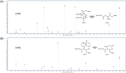
Glutaric aciduria type 1 (GA1) is an inherited neurometabolic disorder, in which deficiency of glutaryl-CoA dehydrogenase leads to accumulation of glutaric acid (GA) and 3-hydroxyglutaric acid (3-HG). Some low excretors may exhibit only slight elevation of urinary 3-HG, with normal urinary GA, yet are at significant risk of severe clinical disease. Accurate quantitation of urinary 3-HG is crucial in diagnostic workup for GA1, but in this context, current gas chromatography–mass spectrometry (GC–MS) methods have inherent analytical challenges. Co-elution and spectral similarities of the 3-HG and 2-HG structural isomers can cause difficulties in quantitation of slightly elevated 3-HG. Our laboratory recently acquired a gas chromatography system coupled to a triple quadrupole mass spectrometer (GC–MS/MS), and we took advantage of its increased sensitivity and specificity to improve our existing GC–MS method. A stable isotope dilution process is used, with sample treatment consisting of a double liquid–liquid extraction followed by a trimethylsilyl derivatization. The transitions m/z 349 → 333 for 3-HG and m/z 349 → 321 for 2-HG were selected to differentiate these two isobaric molecules based on their characteristic fragments, thus minimizing interferences despite co-elution. Method validation demonstrated satisfactory precision and accuracy. Using GC–MS/MS instead of GC–MS allowed us to decrease the required specimen volume, number of sample processing steps, chromatographic run time, and instrument maintenance. This enhanced assay facilitates clinical laboratory testing for GA1, both in confirmatory protocols following positive newborn screening and in diagnostic investigation of patients with suggestive signs or symptoms.

 求助内容:
求助内容: 应助结果提醒方式:


