Peng Li, F. N. U. Alnoor, Wei Xie, Margaret Williams, Julie Feusier, Yi Ding, Xiangrong Zhao, Gang Zheng, Chen Zhao, Arthur W. Zieske, Youli Zu, Philipp W. Raess, Srinivas Tantravahi, Afaf Osman, Ami B. Patel, Tsewang Tashi, Jay L. Patel, Anna P. Matynia, Madhu P. Menon, Rodney R. Miles, Jeffrey R. Jacobsen, Tracy I. George, Douglas W. Sborov, Philippe Szankasi, Paul Rindler, Devin Close, Robert S. Ohgami
{"title":"获得性 UBA1 突变的快速增长使男性患者易患低风险 MDS","authors":"Peng Li, F. N. U. Alnoor, Wei Xie, Margaret Williams, Julie Feusier, Yi Ding, Xiangrong Zhao, Gang Zheng, Chen Zhao, Arthur W. Zieske, Youli Zu, Philipp W. Raess, Srinivas Tantravahi, Afaf Osman, Ami B. Patel, Tsewang Tashi, Jay L. Patel, Anna P. Matynia, Madhu P. Menon, Rodney R. Miles, Jeffrey R. Jacobsen, Tracy I. George, Douglas W. Sborov, Philippe Szankasi, Paul Rindler, Devin Close, Robert S. Ohgami","doi":"10.1038/s41375-024-02397-2","DOIUrl":null,"url":null,"abstract":"<p>Between June 2022 and November 2023, targeted next-generation sequencing (NGS) was performed on blood or bone marrow samples at four US medical centers. We identified 27 distinct presumably somatic <i>UBA1</i> variants in 86 patients (1%, Fig. 1A). Sixty-six patients (0.7%) carried nine different pathogenic/likely pathogenic variants (PV, Fig. 1B above the protein sequence). Most were canonical loss-of-start-codon variants: p.M41T (N = 24), p.M41L (N = 21), and p.M41V (N = 14), followed by previously reported (c.118-1 G>C, N = 2) and two novel splice site variants (c.118-10_118-1 del and c.118-5_118-1 del) upstream of the p.M41 codon. Further, three VEXAS-causal missense variants p.Y55H (N = 1), p.G477A (N = 1), and p.A478S (N = 1) were also classified as PV [1, 2, 4]. An additional 18 distinct novel variants (below the protein sequence in Fig. 1B and in Supplementary Table 1), classified as variants of uncertain significance (VUS), including two recurrent variants (p.D506N and p.I890F), were identified in the remaining 20 patients (Fig. 1B, C, patients 67-86).</p><p>Thirty-one (47%) patients with <i>UBA1</i> PV exhibited at least one concomitant variant, representing a significantly lower frequency compared to VUS patients (85%, p = 0.04, Fig. 1C, D and Table 1), accompanied by a lower somatic mutation burden, defined as the number of somatic variants per patient (Fig. 1E, mean ± SEM, 2.0 ± 0.2 in PV vs. 4.0 ± 0.5 in VUS, p = 0.0001). <i>UBA1</i> clone sizes were notably larger in PV (Fig. 1F, mean ± SEM, 26.1% ± 1.5) than those in VUS (16.0% ± 3.3, p = 0.002). Fifty-five PV patients (83%) exhibited <i>UBA1</i> variant VAFs higher than those of the leading concurrent variants, if any, indicating that <i>UBA1</i> PV were the founding clones. In contrast, only 40% of VUS (Fig. 1G, p = 0.001) were the leading clones. In PV patients, <i>DNMT3A</i> was the most commonly mutated gene (23%), followed by <i>TET2</i> (12%) and <i>ASXL1</i> (6%, Fig. 1C). In VUS patients, the most prevalent concomitant variant was <i>TET2</i> (Supplementary Fig. 1A, 35%, p = 0.03), followed by <i>ASXL1</i> (25%, p = 0.02) and <i>DNMT3A</i> variants (10%, p = 0.03). Notably, variants involved in tyrosine kinase or RAS signaling pathways were significantly more prevalent in VUS patients (Supplementary Fig. 1A–C).</p><figure><figcaption><b data-test=\"table-caption\">Table 1 The clinical diagnosis, molecular and cytogenetic profiles of 86 individuals with presumed somatic UBA1 variants.</b></figcaption><span>Full size table</span><svg aria-hidden=\"true\" focusable=\"false\" height=\"16\" role=\"img\" width=\"16\"><use xlink:href=\"#icon-eds-i-chevron-right-small\" xmlns:xlink=\"http://www.w3.org/1999/xlink\"></use></svg></figure>","PeriodicalId":18109,"journal":{"name":"Leukemia","volume":null,"pages":null},"PeriodicalIF":12.8000,"publicationDate":"2024-11-08","publicationTypes":"Journal Article","fieldsOfStudy":null,"isOpenAccess":false,"openAccessPdf":"","citationCount":"0","resultStr":"{\"title\":\"Rapid growth of acquired UBA1 mutations predisposes male patients to low-risk MDS\",\"authors\":\"Peng Li, F. N. U. Alnoor, Wei Xie, Margaret Williams, Julie Feusier, Yi Ding, Xiangrong Zhao, Gang Zheng, Chen Zhao, Arthur W. Zieske, Youli Zu, Philipp W. Raess, Srinivas Tantravahi, Afaf Osman, Ami B. Patel, Tsewang Tashi, Jay L. Patel, Anna P. Matynia, Madhu P. Menon, Rodney R. Miles, Jeffrey R. Jacobsen, Tracy I. George, Douglas W. Sborov, Philippe Szankasi, Paul Rindler, Devin Close, Robert S. Ohgami\",\"doi\":\"10.1038/s41375-024-02397-2\",\"DOIUrl\":null,\"url\":null,\"abstract\":\"<p>Between June 2022 and November 2023, targeted next-generation sequencing (NGS) was performed on blood or bone marrow samples at four US medical centers. We identified 27 distinct presumably somatic <i>UBA1</i> variants in 86 patients (1%, Fig. 1A). Sixty-six patients (0.7%) carried nine different pathogenic/likely pathogenic variants (PV, Fig. 1B above the protein sequence). Most were canonical loss-of-start-codon variants: p.M41T (N = 24), p.M41L (N = 21), and p.M41V (N = 14), followed by previously reported (c.118-1 G>C, N = 2) and two novel splice site variants (c.118-10_118-1 del and c.118-5_118-1 del) upstream of the p.M41 codon. Further, three VEXAS-causal missense variants p.Y55H (N = 1), p.G477A (N = 1), and p.A478S (N = 1) were also classified as PV [1, 2, 4]. An additional 18 distinct novel variants (below the protein sequence in Fig. 1B and in Supplementary Table 1), classified as variants of uncertain significance (VUS), including two recurrent variants (p.D506N and p.I890F), were identified in the remaining 20 patients (Fig. 1B, C, patients 67-86).</p><p>Thirty-one (47%) patients with <i>UBA1</i> PV exhibited at least one concomitant variant, representing a significantly lower frequency compared to VUS patients (85%, p = 0.04, Fig. 1C, D and Table 1), accompanied by a lower somatic mutation burden, defined as the number of somatic variants per patient (Fig. 1E, mean ± SEM, 2.0 ± 0.2 in PV vs. 4.0 ± 0.5 in VUS, p = 0.0001). <i>UBA1</i> clone sizes were notably larger in PV (Fig. 1F, mean ± SEM, 26.1% ± 1.5) than those in VUS (16.0% ± 3.3, p = 0.002). Fifty-five PV patients (83%) exhibited <i>UBA1</i> variant VAFs higher than those of the leading concurrent variants, if any, indicating that <i>UBA1</i> PV were the founding clones. In contrast, only 40% of VUS (Fig. 1G, p = 0.001) were the leading clones. In PV patients, <i>DNMT3A</i> was the most commonly mutated gene (23%), followed by <i>TET2</i> (12%) and <i>ASXL1</i> (6%, Fig. 1C). In VUS patients, the most prevalent concomitant variant was <i>TET2</i> (Supplementary Fig. 1A, 35%, p = 0.03), followed by <i>ASXL1</i> (25%, p = 0.02) and <i>DNMT3A</i> variants (10%, p = 0.03). Notably, variants involved in tyrosine kinase or RAS signaling pathways were significantly more prevalent in VUS patients (Supplementary Fig. 1A–C).</p><figure><figcaption><b data-test=\\\"table-caption\\\">Table 1 The clinical diagnosis, molecular and cytogenetic profiles of 86 individuals with presumed somatic UBA1 variants.</b></figcaption><span>Full size table</span><svg aria-hidden=\\\"true\\\" focusable=\\\"false\\\" height=\\\"16\\\" role=\\\"img\\\" width=\\\"16\\\"><use xlink:href=\\\"#icon-eds-i-chevron-right-small\\\" xmlns:xlink=\\\"http://www.w3.org/1999/xlink\\\"></use></svg></figure>\",\"PeriodicalId\":18109,\"journal\":{\"name\":\"Leukemia\",\"volume\":null,\"pages\":null},\"PeriodicalIF\":12.8000,\"publicationDate\":\"2024-11-08\",\"publicationTypes\":\"Journal Article\",\"fieldsOfStudy\":null,\"isOpenAccess\":false,\"openAccessPdf\":\"\",\"citationCount\":\"0\",\"resultStr\":null,\"platform\":\"Semanticscholar\",\"paperid\":null,\"PeriodicalName\":\"Leukemia\",\"FirstCategoryId\":\"3\",\"ListUrlMain\":\"https://doi.org/10.1038/s41375-024-02397-2\",\"RegionNum\":1,\"RegionCategory\":\"医学\",\"ArticlePicture\":[],\"TitleCN\":null,\"AbstractTextCN\":null,\"PMCID\":null,\"EPubDate\":\"\",\"PubModel\":\"\",\"JCR\":\"Q1\",\"JCRName\":\"HEMATOLOGY\",\"Score\":null,\"Total\":0}","platform":"Semanticscholar","paperid":null,"PeriodicalName":"Leukemia","FirstCategoryId":"3","ListUrlMain":"https://doi.org/10.1038/s41375-024-02397-2","RegionNum":1,"RegionCategory":"医学","ArticlePicture":[],"TitleCN":null,"AbstractTextCN":null,"PMCID":null,"EPubDate":"","PubModel":"","JCR":"Q1","JCRName":"HEMATOLOGY","Score":null,"Total":0}
Rapid growth of acquired UBA1 mutations predisposes male patients to low-risk MDS
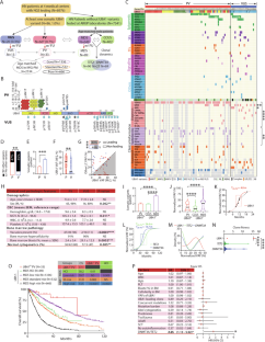
Between June 2022 and November 2023, targeted next-generation sequencing (NGS) was performed on blood or bone marrow samples at four US medical centers. We identified 27 distinct presumably somatic UBA1 variants in 86 patients (1%, Fig. 1A). Sixty-six patients (0.7%) carried nine different pathogenic/likely pathogenic variants (PV, Fig. 1B above the protein sequence). Most were canonical loss-of-start-codon variants: p.M41T (N = 24), p.M41L (N = 21), and p.M41V (N = 14), followed by previously reported (c.118-1 G>C, N = 2) and two novel splice site variants (c.118-10_118-1 del and c.118-5_118-1 del) upstream of the p.M41 codon. Further, three VEXAS-causal missense variants p.Y55H (N = 1), p.G477A (N = 1), and p.A478S (N = 1) were also classified as PV [1, 2, 4]. An additional 18 distinct novel variants (below the protein sequence in Fig. 1B and in Supplementary Table 1), classified as variants of uncertain significance (VUS), including two recurrent variants (p.D506N and p.I890F), were identified in the remaining 20 patients (Fig. 1B, C, patients 67-86).
Thirty-one (47%) patients with UBA1 PV exhibited at least one concomitant variant, representing a significantly lower frequency compared to VUS patients (85%, p = 0.04, Fig. 1C, D and Table 1), accompanied by a lower somatic mutation burden, defined as the number of somatic variants per patient (Fig. 1E, mean ± SEM, 2.0 ± 0.2 in PV vs. 4.0 ± 0.5 in VUS, p = 0.0001). UBA1 clone sizes were notably larger in PV (Fig. 1F, mean ± SEM, 26.1% ± 1.5) than those in VUS (16.0% ± 3.3, p = 0.002). Fifty-five PV patients (83%) exhibited UBA1 variant VAFs higher than those of the leading concurrent variants, if any, indicating that UBA1 PV were the founding clones. In contrast, only 40% of VUS (Fig. 1G, p = 0.001) were the leading clones. In PV patients, DNMT3A was the most commonly mutated gene (23%), followed by TET2 (12%) and ASXL1 (6%, Fig. 1C). In VUS patients, the most prevalent concomitant variant was TET2 (Supplementary Fig. 1A, 35%, p = 0.03), followed by ASXL1 (25%, p = 0.02) and DNMT3A variants (10%, p = 0.03). Notably, variants involved in tyrosine kinase or RAS signaling pathways were significantly more prevalent in VUS patients (Supplementary Fig. 1A–C).
期刊介绍:
Title: Leukemia
Journal Overview:
Publishes high-quality, peer-reviewed research
Covers all aspects of research and treatment of leukemia and allied diseases
Includes studies of normal hemopoiesis due to comparative relevance
Topics of Interest:
Oncogenes
Growth factors
Stem cells
Leukemia genomics
Cell cycle
Signal transduction
Molecular targets for therapy
And more
Content Types:
Original research articles
Reviews
Letters
Correspondence
Comments elaborating on significant advances and covering topical issues