Ting Zhao, Thomas Denize, Hanzhang Wang, Adam S. Fisch, Shulin Wu, Chin-Lee Wu, Kristine M. Cornejo
{"title":"多核肿瘤细胞和微乳头状形态似乎是具有乳头状和肿瘤细胞特征的肾细胞癌预后不良的预测因素。","authors":"Ting Zhao, Thomas Denize, Hanzhang Wang, Adam S. Fisch, Shulin Wu, Chin-Lee Wu, Kristine M. Cornejo","doi":"10.1016/j.humpath.2024.105677","DOIUrl":null,"url":null,"abstract":"<div><div>Renal cell carcinoma with papillary and oncocytic features (RCC-PO) are poorly understood, partially due to conflicting results in multiple studies. The histological features that predict behavior of RCC-PO have not been elucidated. The aim is to review clinicopathologic features and to correlate clinical outcomes of patients with RCC-PO to further expand our knowledge on these heterogeneous tumors. An archival search was done for “RCC” and “papillary,” and tumors with >50% papillary and oncocytic features were included. Clinicopathologic data including tumor size, grade, stage, molecular and immunohistochemical testing when performed, and follow-up data were collected. Using multivariate analyses, correlation between histological features, tumor stage and prognosis were analyzed. Sixty-one patients with RCC-PO were identified of which 49 (80%) were male with a median age of 65 (range: 36–93) years, and a mean tumor size of 5.2 (range: 1–21.5) cm. Micropapillary features were seen in 4, bizarre nuclei (at least 3 times larger or with irregular shape) in 6, multinucleated tumor cells (MTC) in 15, single or small clusters (SSC) (made of 2–3 tumor cells) located away from areas of necrosis in 16, and striking eosinophilic cytoplasmic inclusions in 3 tumors, respectively. Thirty-six (59%) tumors were high-grade (WHO/ISUP grade 3–4), and 23 (38%) had a high stage (≥pT3 or pN1). Tumors were positive for AMACR (15/16) and CK7 (13/17), with preserved FH (7/7) staining and were all negative for CD117 (0/7), ALK, TFE3, cathepsin K, Melan A, and HMB45 (0/4, each). Three tumors underwent chromosomal microarray (CMA) plus gene fusion assay, and FISH and germline testing for <em>FLCN</em> and <em>MET</em> gene alterations by PCR were done on 1 each. Ten (16%) patients had a local recurrence (LR) or metastasis after nephrectomy; 4 died of disease (2 had tumors with micropapillary features), with a median follow-up of 7 (range: 0.01–19) years. Tumors with micropapillary features showed significantly higher RCC-PO-related mortality (50% vs. 3.5%, p < 0.001). In multivariable analysis, SSC correlated with a higher stage (HR: 11.95; p = 0.005); micropapillary features (HR: 18.42; p = 0.017) and MTC (HR: 180.22; p = 0.036) with presence of metastasis/LR; and micropapillary features with a higher RCC-PO-related mortality (HR: 60.35; p = 0.036). RCC-PO are cytogenetically heterogeneous with overlapping features of various renal neoplasms. Micropapillary features and MTC appear to be independent predictors of poor outcomes in these tumors.</div></div>","PeriodicalId":13062,"journal":{"name":"Human pathology","volume":"153 ","pages":"Article 105677"},"PeriodicalIF":2.7000,"publicationDate":"2024-11-01","publicationTypes":"Journal Article","fieldsOfStudy":null,"isOpenAccess":false,"openAccessPdf":"","citationCount":"0","resultStr":"{\"title\":\"Multinucleated tumor cells and micropapillary morphology appear to be predictors of poor prognosis in renal cell carcinoma with papillary and oncocytic features\",\"authors\":\"Ting Zhao, Thomas Denize, Hanzhang Wang, Adam S. Fisch, Shulin Wu, Chin-Lee Wu, Kristine M. Cornejo\",\"doi\":\"10.1016/j.humpath.2024.105677\",\"DOIUrl\":null,\"url\":null,\"abstract\":\"<div><div>Renal cell carcinoma with papillary and oncocytic features (RCC-PO) are poorly understood, partially due to conflicting results in multiple studies. The histological features that predict behavior of RCC-PO have not been elucidated. The aim is to review clinicopathologic features and to correlate clinical outcomes of patients with RCC-PO to further expand our knowledge on these heterogeneous tumors. An archival search was done for “RCC” and “papillary,” and tumors with >50% papillary and oncocytic features were included. Clinicopathologic data including tumor size, grade, stage, molecular and immunohistochemical testing when performed, and follow-up data were collected. Using multivariate analyses, correlation between histological features, tumor stage and prognosis were analyzed. Sixty-one patients with RCC-PO were identified of which 49 (80%) were male with a median age of 65 (range: 36–93) years, and a mean tumor size of 5.2 (range: 1–21.5) cm. Micropapillary features were seen in 4, bizarre nuclei (at least 3 times larger or with irregular shape) in 6, multinucleated tumor cells (MTC) in 15, single or small clusters (SSC) (made of 2–3 tumor cells) located away from areas of necrosis in 16, and striking eosinophilic cytoplasmic inclusions in 3 tumors, respectively. Thirty-six (59%) tumors were high-grade (WHO/ISUP grade 3–4), and 23 (38%) had a high stage (≥pT3 or pN1). Tumors were positive for AMACR (15/16) and CK7 (13/17), with preserved FH (7/7) staining and were all negative for CD117 (0/7), ALK, TFE3, cathepsin K, Melan A, and HMB45 (0/4, each). Three tumors underwent chromosomal microarray (CMA) plus gene fusion assay, and FISH and germline testing for <em>FLCN</em> and <em>MET</em> gene alterations by PCR were done on 1 each. Ten (16%) patients had a local recurrence (LR) or metastasis after nephrectomy; 4 died of disease (2 had tumors with micropapillary features), with a median follow-up of 7 (range: 0.01–19) years. Tumors with micropapillary features showed significantly higher RCC-PO-related mortality (50% vs. 3.5%, p < 0.001). In multivariable analysis, SSC correlated with a higher stage (HR: 11.95; p = 0.005); micropapillary features (HR: 18.42; p = 0.017) and MTC (HR: 180.22; p = 0.036) with presence of metastasis/LR; and micropapillary features with a higher RCC-PO-related mortality (HR: 60.35; p = 0.036). RCC-PO are cytogenetically heterogeneous with overlapping features of various renal neoplasms. Micropapillary features and MTC appear to be independent predictors of poor outcomes in these tumors.</div></div>\",\"PeriodicalId\":13062,\"journal\":{\"name\":\"Human pathology\",\"volume\":\"153 \",\"pages\":\"Article 105677\"},\"PeriodicalIF\":2.7000,\"publicationDate\":\"2024-11-01\",\"publicationTypes\":\"Journal Article\",\"fieldsOfStudy\":null,\"isOpenAccess\":false,\"openAccessPdf\":\"\",\"citationCount\":\"0\",\"resultStr\":null,\"platform\":\"Semanticscholar\",\"paperid\":null,\"PeriodicalName\":\"Human pathology\",\"FirstCategoryId\":\"3\",\"ListUrlMain\":\"https://www.sciencedirect.com/science/article/pii/S0046817724001862\",\"RegionNum\":2,\"RegionCategory\":\"医学\",\"ArticlePicture\":[],\"TitleCN\":null,\"AbstractTextCN\":null,\"PMCID\":null,\"EPubDate\":\"\",\"PubModel\":\"\",\"JCR\":\"Q2\",\"JCRName\":\"PATHOLOGY\",\"Score\":null,\"Total\":0}","platform":"Semanticscholar","paperid":null,"PeriodicalName":"Human pathology","FirstCategoryId":"3","ListUrlMain":"https://www.sciencedirect.com/science/article/pii/S0046817724001862","RegionNum":2,"RegionCategory":"医学","ArticlePicture":[],"TitleCN":null,"AbstractTextCN":null,"PMCID":null,"EPubDate":"","PubModel":"","JCR":"Q2","JCRName":"PATHOLOGY","Score":null,"Total":0}
Multinucleated tumor cells and micropapillary morphology appear to be predictors of poor prognosis in renal cell carcinoma with papillary and oncocytic features
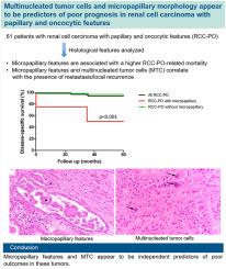
Renal cell carcinoma with papillary and oncocytic features (RCC-PO) are poorly understood, partially due to conflicting results in multiple studies. The histological features that predict behavior of RCC-PO have not been elucidated. The aim is to review clinicopathologic features and to correlate clinical outcomes of patients with RCC-PO to further expand our knowledge on these heterogeneous tumors. An archival search was done for “RCC” and “papillary,” and tumors with >50% papillary and oncocytic features were included. Clinicopathologic data including tumor size, grade, stage, molecular and immunohistochemical testing when performed, and follow-up data were collected. Using multivariate analyses, correlation between histological features, tumor stage and prognosis were analyzed. Sixty-one patients with RCC-PO were identified of which 49 (80%) were male with a median age of 65 (range: 36–93) years, and a mean tumor size of 5.2 (range: 1–21.5) cm. Micropapillary features were seen in 4, bizarre nuclei (at least 3 times larger or with irregular shape) in 6, multinucleated tumor cells (MTC) in 15, single or small clusters (SSC) (made of 2–3 tumor cells) located away from areas of necrosis in 16, and striking eosinophilic cytoplasmic inclusions in 3 tumors, respectively. Thirty-six (59%) tumors were high-grade (WHO/ISUP grade 3–4), and 23 (38%) had a high stage (≥pT3 or pN1). Tumors were positive for AMACR (15/16) and CK7 (13/17), with preserved FH (7/7) staining and were all negative for CD117 (0/7), ALK, TFE3, cathepsin K, Melan A, and HMB45 (0/4, each). Three tumors underwent chromosomal microarray (CMA) plus gene fusion assay, and FISH and germline testing for FLCN and MET gene alterations by PCR were done on 1 each. Ten (16%) patients had a local recurrence (LR) or metastasis after nephrectomy; 4 died of disease (2 had tumors with micropapillary features), with a median follow-up of 7 (range: 0.01–19) years. Tumors with micropapillary features showed significantly higher RCC-PO-related mortality (50% vs. 3.5%, p < 0.001). In multivariable analysis, SSC correlated with a higher stage (HR: 11.95; p = 0.005); micropapillary features (HR: 18.42; p = 0.017) and MTC (HR: 180.22; p = 0.036) with presence of metastasis/LR; and micropapillary features with a higher RCC-PO-related mortality (HR: 60.35; p = 0.036). RCC-PO are cytogenetically heterogeneous with overlapping features of various renal neoplasms. Micropapillary features and MTC appear to be independent predictors of poor outcomes in these tumors.
期刊介绍:
Human Pathology is designed to bring information of clinicopathologic significance to human disease to the laboratory and clinical physician. It presents information drawn from morphologic and clinical laboratory studies with direct relevance to the understanding of human diseases. Papers published concern morphologic and clinicopathologic observations, reviews of diseases, analyses of problems in pathology, significant collections of case material and advances in concepts or techniques of value in the analysis and diagnosis of disease. Theoretical and experimental pathology and molecular biology pertinent to human disease are included. This critical journal is well illustrated with exceptional reproductions of photomicrographs and microscopic anatomy.