Camilla Merrild , Gitte A. Pedersen , Kristian W. Antonsen , Mia G. Madsen , Anna K. Keller , Holger J. Møller , Lene N. Nejsum , Henricus A.M. Mutsaers , Rikke Nørregaard
{"title":"基于人体组织的肾脏炎症模型","authors":"Camilla Merrild , Gitte A. Pedersen , Kristian W. Antonsen , Mia G. Madsen , Anna K. Keller , Holger J. Møller , Lene N. Nejsum , Henricus A.M. Mutsaers , Rikke Nørregaard","doi":"10.1016/j.yexcr.2024.114309","DOIUrl":null,"url":null,"abstract":"<div><div>Inflammation plays a key role in both the onset and progression of various kidney diseases. However, the specific molecular and cellular mechanisms by which inflammation drives kidney diseases from different etiologies remain to be elucidated. To enhance our understanding of these mechanisms, a reliable and translational human model of renal inflammation is needed.</div><div>Here, we aim to establish such a model using human precision-cut kidney slices (PCKS). The PCKS were prepared from fresh, macroscopically healthy kidney tissue and cultured for 3h–48h with or without tumor necrosis factor-α (TNFα), or its inhibitor Etanercept. The ensuing inflammatory response in the slices was evaluated using both qPCR and a cytokine array. Furthermore, the presence of immune cells was visualized using immunofluorescent staining, and the activation potential of tissue-resident macrophages was examined with ELISA.</div><div>We observed a culture-induced inflammatory response, reflected by increased expression of pro-inflammatory genes <em>TNF</em>, <em>IL1B</em>, <em>CCL2</em>, and <em>IL6</em>. This response could be partially inhibited by Etanercept, indicating that TNFα plays a role in the observed response. Moreover, we found that TNFα stimulation further increased the gene expression of <em>TNF</em>, <em>IL1B</em>, <em>CCL2</em>, and <em>IL6</em>, as well as the production of several chemokines and cytokines, including CXCL5, MCP1, MCP3, and IL-6. Lastly, we observed the presence of CD14- and HLA-DR-positive cells, as well as proliferating (CD68- and PCNA-positive) and activated macrophages in the slices during incubation. In conclusion, this study presents a novel human model for investigating renal inflammation.</div></div>","PeriodicalId":12227,"journal":{"name":"Experimental cell research","volume":"443 1","pages":"Article 114309"},"PeriodicalIF":3.3000,"publicationDate":"2024-11-01","publicationTypes":"Journal Article","fieldsOfStudy":null,"isOpenAccess":false,"openAccessPdf":"","citationCount":"0","resultStr":"{\"title\":\"A human tissue-based model of renal inflammation\",\"authors\":\"Camilla Merrild , Gitte A. Pedersen , Kristian W. Antonsen , Mia G. Madsen , Anna K. Keller , Holger J. Møller , Lene N. Nejsum , Henricus A.M. Mutsaers , Rikke Nørregaard\",\"doi\":\"10.1016/j.yexcr.2024.114309\",\"DOIUrl\":null,\"url\":null,\"abstract\":\"<div><div>Inflammation plays a key role in both the onset and progression of various kidney diseases. However, the specific molecular and cellular mechanisms by which inflammation drives kidney diseases from different etiologies remain to be elucidated. To enhance our understanding of these mechanisms, a reliable and translational human model of renal inflammation is needed.</div><div>Here, we aim to establish such a model using human precision-cut kidney slices (PCKS). The PCKS were prepared from fresh, macroscopically healthy kidney tissue and cultured for 3h–48h with or without tumor necrosis factor-α (TNFα), or its inhibitor Etanercept. The ensuing inflammatory response in the slices was evaluated using both qPCR and a cytokine array. Furthermore, the presence of immune cells was visualized using immunofluorescent staining, and the activation potential of tissue-resident macrophages was examined with ELISA.</div><div>We observed a culture-induced inflammatory response, reflected by increased expression of pro-inflammatory genes <em>TNF</em>, <em>IL1B</em>, <em>CCL2</em>, and <em>IL6</em>. This response could be partially inhibited by Etanercept, indicating that TNFα plays a role in the observed response. Moreover, we found that TNFα stimulation further increased the gene expression of <em>TNF</em>, <em>IL1B</em>, <em>CCL2</em>, and <em>IL6</em>, as well as the production of several chemokines and cytokines, including CXCL5, MCP1, MCP3, and IL-6. Lastly, we observed the presence of CD14- and HLA-DR-positive cells, as well as proliferating (CD68- and PCNA-positive) and activated macrophages in the slices during incubation. In conclusion, this study presents a novel human model for investigating renal inflammation.</div></div>\",\"PeriodicalId\":12227,\"journal\":{\"name\":\"Experimental cell research\",\"volume\":\"443 1\",\"pages\":\"Article 114309\"},\"PeriodicalIF\":3.3000,\"publicationDate\":\"2024-11-01\",\"publicationTypes\":\"Journal Article\",\"fieldsOfStudy\":null,\"isOpenAccess\":false,\"openAccessPdf\":\"\",\"citationCount\":\"0\",\"resultStr\":null,\"platform\":\"Semanticscholar\",\"paperid\":null,\"PeriodicalName\":\"Experimental cell research\",\"FirstCategoryId\":\"3\",\"ListUrlMain\":\"https://www.sciencedirect.com/science/article/pii/S0014482724004002\",\"RegionNum\":3,\"RegionCategory\":\"生物学\",\"ArticlePicture\":[],\"TitleCN\":null,\"AbstractTextCN\":null,\"PMCID\":null,\"EPubDate\":\"\",\"PubModel\":\"\",\"JCR\":\"Q3\",\"JCRName\":\"CELL BIOLOGY\",\"Score\":null,\"Total\":0}","platform":"Semanticscholar","paperid":null,"PeriodicalName":"Experimental cell research","FirstCategoryId":"3","ListUrlMain":"https://www.sciencedirect.com/science/article/pii/S0014482724004002","RegionNum":3,"RegionCategory":"生物学","ArticlePicture":[],"TitleCN":null,"AbstractTextCN":null,"PMCID":null,"EPubDate":"","PubModel":"","JCR":"Q3","JCRName":"CELL BIOLOGY","Score":null,"Total":0}
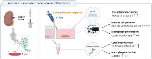
Inflammation plays a key role in both the onset and progression of various kidney diseases. However, the specific molecular and cellular mechanisms by which inflammation drives kidney diseases from different etiologies remain to be elucidated. To enhance our understanding of these mechanisms, a reliable and translational human model of renal inflammation is needed.
Here, we aim to establish such a model using human precision-cut kidney slices (PCKS). The PCKS were prepared from fresh, macroscopically healthy kidney tissue and cultured for 3h–48h with or without tumor necrosis factor-α (TNFα), or its inhibitor Etanercept. The ensuing inflammatory response in the slices was evaluated using both qPCR and a cytokine array. Furthermore, the presence of immune cells was visualized using immunofluorescent staining, and the activation potential of tissue-resident macrophages was examined with ELISA.
We observed a culture-induced inflammatory response, reflected by increased expression of pro-inflammatory genes TNF, IL1B, CCL2, and IL6. This response could be partially inhibited by Etanercept, indicating that TNFα plays a role in the observed response. Moreover, we found that TNFα stimulation further increased the gene expression of TNF, IL1B, CCL2, and IL6, as well as the production of several chemokines and cytokines, including CXCL5, MCP1, MCP3, and IL-6. Lastly, we observed the presence of CD14- and HLA-DR-positive cells, as well as proliferating (CD68- and PCNA-positive) and activated macrophages in the slices during incubation. In conclusion, this study presents a novel human model for investigating renal inflammation.
期刊介绍:
Our scope includes but is not limited to areas such as: Chromosome biology; Chromatin and epigenetics; DNA repair; Gene regulation; Nuclear import-export; RNA processing; Non-coding RNAs; Organelle biology; The cytoskeleton; Intracellular trafficking; Cell-cell and cell-matrix interactions; Cell motility and migration; Cell proliferation; Cellular differentiation; Signal transduction; Programmed cell death.