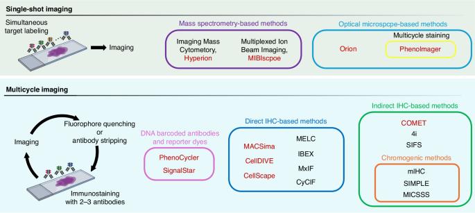
利用当前的多重成像技术进行空间分析,以确定癌症组织的分子特征。

IF 6.4 1区 医学 Q1 ONCOLOGY
Takashi Semba, Takatsugu Ishimoto
{"title":"利用当前的多重成像技术进行空间分析,以确定癌症组织的分子特征。","authors":"Takashi Semba, Takatsugu Ishimoto","doi":"10.1038/s41416-024-02882-6","DOIUrl":null,"url":null,"abstract":"<p><p>Tumours are composed of tumour cells and the surrounding tumour microenvironment (TME), and the molecular characterisation of the various elements of the TME and their interactions is essential for elucidating the mechanisms of tumour progression and developing better therapeutic strategies. Multiplex imaging is a technique that can quantify the expression of multiple protein markers on the same tissue section while maintaining spatial positioning, and this method has been rapidly developed in cancer research in recent years. Many multiplex imaging technologies and spatial analysis methods are emerging, and the elucidation of their principles and features is essential. In this review, we provide an overview of the latest multiplex imaging techniques by type of imaging and staining method and an introduction to image analysis methods, primarily focusing on spatial cellular properties, providing deeper insight into tumour organisation and spatial molecular biology in the TME.</p>","PeriodicalId":9243,"journal":{"name":"British Journal of Cancer","volume":" ","pages":""},"PeriodicalIF":6.4000,"publicationDate":"2024-10-22","publicationTypes":"Journal Article","fieldsOfStudy":null,"isOpenAccess":false,"openAccessPdf":"","citationCount":"0","resultStr":"{\"title\":\"Spatial analysis by current multiplexed imaging technologies for the molecular characterisation of cancer tissues.\",\"authors\":\"Takashi Semba, Takatsugu Ishimoto\",\"doi\":\"10.1038/s41416-024-02882-6\",\"DOIUrl\":null,\"url\":null,\"abstract\":\"<p><p>Tumours are composed of tumour cells and the surrounding tumour microenvironment (TME), and the molecular characterisation of the various elements of the TME and their interactions is essential for elucidating the mechanisms of tumour progression and developing better therapeutic strategies. Multiplex imaging is a technique that can quantify the expression of multiple protein markers on the same tissue section while maintaining spatial positioning, and this method has been rapidly developed in cancer research in recent years. Many multiplex imaging technologies and spatial analysis methods are emerging, and the elucidation of their principles and features is essential. In this review, we provide an overview of the latest multiplex imaging techniques by type of imaging and staining method and an introduction to image analysis methods, primarily focusing on spatial cellular properties, providing deeper insight into tumour organisation and spatial molecular biology in the TME.</p>\",\"PeriodicalId\":9243,\"journal\":{\"name\":\"British Journal of Cancer\",\"volume\":\" \",\"pages\":\"\"},\"PeriodicalIF\":6.4000,\"publicationDate\":\"2024-10-22\",\"publicationTypes\":\"Journal Article\",\"fieldsOfStudy\":null,\"isOpenAccess\":false,\"openAccessPdf\":\"\",\"citationCount\":\"0\",\"resultStr\":null,\"platform\":\"Semanticscholar\",\"paperid\":null,\"PeriodicalName\":\"British Journal of Cancer\",\"FirstCategoryId\":\"3\",\"ListUrlMain\":\"https://doi.org/10.1038/s41416-024-02882-6\",\"RegionNum\":1,\"RegionCategory\":\"医学\",\"ArticlePicture\":[],\"TitleCN\":null,\"AbstractTextCN\":null,\"PMCID\":null,\"EPubDate\":\"\",\"PubModel\":\"\",\"JCR\":\"Q1\",\"JCRName\":\"ONCOLOGY\",\"Score\":null,\"Total\":0}","platform":"Semanticscholar","paperid":null,"PeriodicalName":"British Journal of Cancer","FirstCategoryId":"3","ListUrlMain":"https://doi.org/10.1038/s41416-024-02882-6","RegionNum":1,"RegionCategory":"医学","ArticlePicture":[],"TitleCN":null,"AbstractTextCN":null,"PMCID":null,"EPubDate":"","PubModel":"","JCR":"Q1","JCRName":"ONCOLOGY","Score":null,"Total":0}
引用次数: 0

摘要

肿瘤由肿瘤细胞和周围的肿瘤微环境(TME)组成,TME 各要素的分子特征及其相互作用对于阐明肿瘤进展机制和开发更好的治疗策略至关重要。多重成像是一种能在保持空间定位的情况下量化同一组织切片上多种蛋白质标记物表达的技术,这种方法近年来在癌症研究领域得到了迅速发展。许多多重成像技术和空间分析方法不断涌现,阐明其原理和特点至关重要。在这篇综述中,我们按成像和染色方法的类型概述了最新的多重成像技术,并介绍了主要侧重于细胞空间特性的图像分析方法,从而为深入了解肿瘤组织和 TME 中的空间分子生物学提供了依据。
本文章由计算机程序翻译,如有差异,请以英文原文为准。
Spatial analysis by current multiplexed imaging technologies for the molecular characterisation of cancer tissues.

Tumours are composed of tumour cells and the surrounding tumour microenvironment (TME), and the molecular characterisation of the various elements of the TME and their interactions is essential for elucidating the mechanisms of tumour progression and developing better therapeutic strategies. Multiplex imaging is a technique that can quantify the expression of multiple protein markers on the same tissue section while maintaining spatial positioning, and this method has been rapidly developed in cancer research in recent years. Many multiplex imaging technologies and spatial analysis methods are emerging, and the elucidation of their principles and features is essential. In this review, we provide an overview of the latest multiplex imaging techniques by type of imaging and staining method and an introduction to image analysis methods, primarily focusing on spatial cellular properties, providing deeper insight into tumour organisation and spatial molecular biology in the TME.

求助全文
通过发布文献求助,成功后即可免费获取论文全文。 去求助
来源期刊
British Journal of Cancer
British Journal of Cancer 医学-肿瘤学
CiteScore
15.10
自引率
1.10%
发文量
383
审稿时长
6 months
期刊介绍: The British Journal of Cancer is one of the most-cited general cancer journals, publishing significant advances in translational and clinical cancer research.It also publishes high-quality reviews and thought-provoking comment on all aspects of cancer prevention,diagnosis and treatment.
×
引用
GB/T 7714-2015
复制
MLA
复制
APA
复制
导出至
BibTeX EndNote RefMan NoteFirst NoteExpress
×
提示
您的信息不完整,为了账户安全,请先补充。
现在去补充
×
提示
您因"违规操作"
具体请查看互助需知
我知道了
×
提示
确定
请完成安全验证×
copy
已复制链接
快去分享给好友吧!
我知道了
右上角分享
点击右上角分享
0
联系我们:info@booksci.cn Book学术提供免费学术资源搜索服务,方便国内外学者检索中英文文献。致力于提供最便捷和优质的服务体验。 Copyright © 2023 布克学术 All rights reserved.
京ICP备2023020795号-1
ghs 京公网安备 11010802042870号
Book学术文献互助
Book学术文献互助群
群 号:481959085
Book学术官方微信