Lan Zhang , Quan-Xiang Cui , Liang-Qin Zhou , Xin-Yi Wang , Hong-Xia Zhang , Yue-Min Zhu , Xi-Qiao Sang , Zi-Xiang Kuai
{"title":"基于磁共振成像的载体放射组学用于预测乳腺癌 HER2 状态及其在新辅助治疗后的变化。","authors":"Lan Zhang , Quan-Xiang Cui , Liang-Qin Zhou , Xin-Yi Wang , Hong-Xia Zhang , Yue-Min Zhu , Xi-Qiao Sang , Zi-Xiang Kuai","doi":"10.1016/j.compmedimag.2024.102443","DOIUrl":null,"url":null,"abstract":"<div><h3>Purpose</h3><div>: To develop a novel MRI-based vector radiomic approach to predict breast cancer (BC) human epidermal growth factor receptor 2 (HER2) status (zero, low, and positive; task 1) and its changes after neoadjuvant therapy (NAT) (positive-to-positive, positive-to-negative, and positive-to-pathologic complete response; task 2).</div></div><div><h3>Materials and Methods</h3><div>: Both dynamic contrast-enhanced (DCE) MRI data and multi-<em>b</em>-value (MBV) diffusion-weighted imaging (DWI) data were acquired in BC patients at two centers. Vector-radiomic and conventional-radiomic features were extracted from both DCE-MRI and MBV-DWI. After feature selection, the following models were built using the retained features and logistic regression: vector model, conventional model, and combined model that integrates the vector-radiomic and conventional-radiomic features. The models’ performances were quantified by the area under the receiver-operating characteristic curve (AUC).</div></div><div><h3>Results:</h3><div>The training/external test set (center 1/2) included 483/361 women. For task 1, the vector model (AUCs=0.73<span><math><mo>∼</mo></math></span>0.86) was superior to (<em>p</em><span><math><mo><</mo></math></span>.05) the conventional model (AUCs=0.68<span><math><mo>∼</mo></math></span>0.81), and the addition of vector-radiomic features to conventional-radiomic features yielded an incremental predictive value (AUCs=0.80<span><math><mo>∼</mo></math></span>0.90, <span><math><mrow><mi>p</mi><mo><</mo><mo>.</mo><mn>05</mn></mrow></math></span>). For task 2, the combined MBV-DWI model (AUCs=0.85<span><math><mo>∼</mo></math></span>0.89) performed better than (<span><math><mrow><mi>p</mi><mo><</mo><mo>.</mo><mn>05</mn></mrow></math></span>) the conventional MBV-DWI model (AUCs=0.73<span><math><mo>∼</mo></math></span>0.82). In addition, for the combined DCE-MRI model and the combined MBV-DWI model, the former (AUCs=0.85<span><math><mo>∼</mo></math></span>0.90) outperformed (<span><math><mrow><mi>p</mi><mo><</mo><mo>.</mo><mn>05</mn></mrow></math></span>) the latter (AUCs=0.80<span><math><mo>∼</mo></math></span>0.85) in task 1, whereas the latter (AUCs=0.85<span><math><mo>∼</mo></math></span>0.89) outperformed (<span><math><mrow><mi>p</mi><mo><</mo><mo>.</mo><mn>05</mn></mrow></math></span>) the former (AUCs=0.76<span><math><mo>∼</mo></math></span>0.81) in task 2. The above results are true for the training and external test sets.</div></div><div><h3>Conclusions:</h3><div>MRI-based vector radiomics may predict BC HER2 status and its changes after NAT and provide significant incremental prediction over and above conventional radiomics.</div></div>","PeriodicalId":50631,"journal":{"name":"Computerized Medical Imaging and Graphics","volume":"118 ","pages":"Article 102443"},"PeriodicalIF":5.4000,"publicationDate":"2024-10-17","publicationTypes":"Journal Article","fieldsOfStudy":null,"isOpenAccess":false,"openAccessPdf":"","citationCount":"0","resultStr":"{\"title\":\"MRI-based vector radiomics for predicting breast cancer HER2 status and its changes after neoadjuvant therapy\",\"authors\":\"Lan Zhang , Quan-Xiang Cui , Liang-Qin Zhou , Xin-Yi Wang , Hong-Xia Zhang , Yue-Min Zhu , Xi-Qiao Sang , Zi-Xiang Kuai\",\"doi\":\"10.1016/j.compmedimag.2024.102443\",\"DOIUrl\":null,\"url\":null,\"abstract\":\"<div><h3>Purpose</h3><div>: To develop a novel MRI-based vector radiomic approach to predict breast cancer (BC) human epidermal growth factor receptor 2 (HER2) status (zero, low, and positive; task 1) and its changes after neoadjuvant therapy (NAT) (positive-to-positive, positive-to-negative, and positive-to-pathologic complete response; task 2).</div></div><div><h3>Materials and Methods</h3><div>: Both dynamic contrast-enhanced (DCE) MRI data and multi-<em>b</em>-value (MBV) diffusion-weighted imaging (DWI) data were acquired in BC patients at two centers. Vector-radiomic and conventional-radiomic features were extracted from both DCE-MRI and MBV-DWI. After feature selection, the following models were built using the retained features and logistic regression: vector model, conventional model, and combined model that integrates the vector-radiomic and conventional-radiomic features. The models’ performances were quantified by the area under the receiver-operating characteristic curve (AUC).</div></div><div><h3>Results:</h3><div>The training/external test set (center 1/2) included 483/361 women. For task 1, the vector model (AUCs=0.73<span><math><mo>∼</mo></math></span>0.86) was superior to (<em>p</em><span><math><mo><</mo></math></span>.05) the conventional model (AUCs=0.68<span><math><mo>∼</mo></math></span>0.81), and the addition of vector-radiomic features to conventional-radiomic features yielded an incremental predictive value (AUCs=0.80<span><math><mo>∼</mo></math></span>0.90, <span><math><mrow><mi>p</mi><mo><</mo><mo>.</mo><mn>05</mn></mrow></math></span>). For task 2, the combined MBV-DWI model (AUCs=0.85<span><math><mo>∼</mo></math></span>0.89) performed better than (<span><math><mrow><mi>p</mi><mo><</mo><mo>.</mo><mn>05</mn></mrow></math></span>) the conventional MBV-DWI model (AUCs=0.73<span><math><mo>∼</mo></math></span>0.82). In addition, for the combined DCE-MRI model and the combined MBV-DWI model, the former (AUCs=0.85<span><math><mo>∼</mo></math></span>0.90) outperformed (<span><math><mrow><mi>p</mi><mo><</mo><mo>.</mo><mn>05</mn></mrow></math></span>) the latter (AUCs=0.80<span><math><mo>∼</mo></math></span>0.85) in task 1, whereas the latter (AUCs=0.85<span><math><mo>∼</mo></math></span>0.89) outperformed (<span><math><mrow><mi>p</mi><mo><</mo><mo>.</mo><mn>05</mn></mrow></math></span>) the former (AUCs=0.76<span><math><mo>∼</mo></math></span>0.81) in task 2. The above results are true for the training and external test sets.</div></div><div><h3>Conclusions:</h3><div>MRI-based vector radiomics may predict BC HER2 status and its changes after NAT and provide significant incremental prediction over and above conventional radiomics.</div></div>\",\"PeriodicalId\":50631,\"journal\":{\"name\":\"Computerized Medical Imaging and Graphics\",\"volume\":\"118 \",\"pages\":\"Article 102443\"},\"PeriodicalIF\":5.4000,\"publicationDate\":\"2024-10-17\",\"publicationTypes\":\"Journal Article\",\"fieldsOfStudy\":null,\"isOpenAccess\":false,\"openAccessPdf\":\"\",\"citationCount\":\"0\",\"resultStr\":null,\"platform\":\"Semanticscholar\",\"paperid\":null,\"PeriodicalName\":\"Computerized Medical Imaging and Graphics\",\"FirstCategoryId\":\"5\",\"ListUrlMain\":\"https://www.sciencedirect.com/science/article/pii/S0895611124001204\",\"RegionNum\":2,\"RegionCategory\":\"医学\",\"ArticlePicture\":[],\"TitleCN\":null,\"AbstractTextCN\":null,\"PMCID\":null,\"EPubDate\":\"\",\"PubModel\":\"\",\"JCR\":\"Q1\",\"JCRName\":\"ENGINEERING, BIOMEDICAL\",\"Score\":null,\"Total\":0}","platform":"Semanticscholar","paperid":null,"PeriodicalName":"Computerized Medical Imaging and Graphics","FirstCategoryId":"5","ListUrlMain":"https://www.sciencedirect.com/science/article/pii/S0895611124001204","RegionNum":2,"RegionCategory":"医学","ArticlePicture":[],"TitleCN":null,"AbstractTextCN":null,"PMCID":null,"EPubDate":"","PubModel":"","JCR":"Q1","JCRName":"ENGINEERING, BIOMEDICAL","Score":null,"Total":0}
引用次数: 0
摘要
目的:开发一种基于磁共振成像的新型矢量放射组学方法,用于预测乳腺癌(BC)人表皮生长因子受体2(HER2)状态(零、低和阳性;任务1)及其在新辅助治疗(NAT)后的变化(阳性到阳性、阳性到阴性、阳性到病理完全反应;任务2):在两个中心采集了 BC 患者的动态对比增强(DCE)磁共振成像数据和多比值(MBV)弥散加权成像(DWI)数据。从 DCE-MRI 和 MBV-DWI 中提取了矢量放射学和传统放射学特征。经过特征选择后,利用保留的特征和逻辑回归建立了以下模型:矢量模型、传统模型以及整合了矢量放射体和传统放射体特征的组合模型。模型的性能通过接收者工作特征曲线下面积(AUC)进行量化:训练/外部测试集(中心 1/2)包括 483/361 名女性。在任务 1 中,矢量模型(AUCs=0.73∼0.86)优于矢量模型(pConclusions:基于 MRI 的矢量放射组学可预测 BC HER2 状态及其在 NAT 后的变化,其预测效果明显优于传统放射组学。
MRI-based vector radiomics for predicting breast cancer HER2 status and its changes after neoadjuvant therapy
Purpose
: To develop a novel MRI-based vector radiomic approach to predict breast cancer (BC) human epidermal growth factor receptor 2 (HER2) status (zero, low, and positive; task 1) and its changes after neoadjuvant therapy (NAT) (positive-to-positive, positive-to-negative, and positive-to-pathologic complete response; task 2).
Materials and Methods
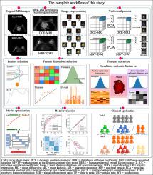
: Both dynamic contrast-enhanced (DCE) MRI data and multi-b-value (MBV) diffusion-weighted imaging (DWI) data were acquired in BC patients at two centers. Vector-radiomic and conventional-radiomic features were extracted from both DCE-MRI and MBV-DWI. After feature selection, the following models were built using the retained features and logistic regression: vector model, conventional model, and combined model that integrates the vector-radiomic and conventional-radiomic features. The models’ performances were quantified by the area under the receiver-operating characteristic curve (AUC).
Results:
The training/external test set (center 1/2) included 483/361 women. For task 1, the vector model (AUCs=0.730.86) was superior to (p.05) the conventional model (AUCs=0.680.81), and the addition of vector-radiomic features to conventional-radiomic features yielded an incremental predictive value (AUCs=0.800.90, ). For task 2, the combined MBV-DWI model (AUCs=0.850.89) performed better than () the conventional MBV-DWI model (AUCs=0.730.82). In addition, for the combined DCE-MRI model and the combined MBV-DWI model, the former (AUCs=0.850.90) outperformed () the latter (AUCs=0.800.85) in task 1, whereas the latter (AUCs=0.850.89) outperformed () the former (AUCs=0.760.81) in task 2. The above results are true for the training and external test sets.
Conclusions:
MRI-based vector radiomics may predict BC HER2 status and its changes after NAT and provide significant incremental prediction over and above conventional radiomics.
期刊介绍:
The purpose of the journal Computerized Medical Imaging and Graphics is to act as a source for the exchange of research results concerning algorithmic advances, development, and application of digital imaging in disease detection, diagnosis, intervention, prevention, precision medicine, and population health. Included in the journal will be articles on novel computerized imaging or visualization techniques, including artificial intelligence and machine learning, augmented reality for surgical planning and guidance, big biomedical data visualization, computer-aided diagnosis, computerized-robotic surgery, image-guided therapy, imaging scanning and reconstruction, mobile and tele-imaging, radiomics, and imaging integration and modeling with other information relevant to digital health. The types of biomedical imaging include: magnetic resonance, computed tomography, ultrasound, nuclear medicine, X-ray, microwave, optical and multi-photon microscopy, video and sensory imaging, and the convergence of biomedical images with other non-imaging datasets.