{"title":"模仿管状先天性食管重复的食管损伤--诊断难题:病例报告。","authors":"Shreya Shrivastava, Priscilla Joshi, Shriyash Pinglikar","doi":"10.1093/bjrcr/uaae035","DOIUrl":null,"url":null,"abstract":"<p><p>Intramural oesophageal dissection (IED) is an uncommon condition in newborns marked by the separation of the mucosal and submucosal layers of the oesophageal wall, both transversely and longitudinally, which may or may not involve perforation. A neonate presented at 26 h of life with poor respiratory effort and lethargy. She was intubated and was put on mechanical ventilation. Radiograph of the neonate suggested malpositioned endotracheal tube. The fluoroscopic dye-study indicated gastroesophageal oesophageal reflux disease and nothing significant. On limited CT contrast study of thorax, a tubular structure was seen running just parallel to the oesophagus extending from the T2 to the T9 levels. Possibilities of a oesophageal duplication/IED were considered. The neonate underwent an endoscopy and gastrostomy on day of life (DOL) 9. On follow up at 3 months a repeat limited CT study was done with instillation of water-soluble contrast. The previously seen tubular structure running parallel to the oesophagus was no longer seen. This finding suggested a healed IED. This case report emphasizes the significance of multimodality imaging in the diagnosis of this condition.</p>","PeriodicalId":45216,"journal":{"name":"BJR Case Reports","volume":"10 5","pages":"uaae035"},"PeriodicalIF":0.5000,"publicationDate":"2024-10-03","publicationTypes":"Journal Article","fieldsOfStudy":null,"isOpenAccess":false,"openAccessPdf":"https://www.ncbi.nlm.nih.gov/pmc/articles/PMC11486540/pdf/","citationCount":"0","resultStr":"{\"title\":\"Oesophageal injury mimicking a tubular congenital oesophageal duplication-a diagnostic dilemma: a case report.\",\"authors\":\"Shreya Shrivastava, Priscilla Joshi, Shriyash Pinglikar\",\"doi\":\"10.1093/bjrcr/uaae035\",\"DOIUrl\":null,\"url\":null,\"abstract\":\"<p><p>Intramural oesophageal dissection (IED) is an uncommon condition in newborns marked by the separation of the mucosal and submucosal layers of the oesophageal wall, both transversely and longitudinally, which may or may not involve perforation. A neonate presented at 26 h of life with poor respiratory effort and lethargy. She was intubated and was put on mechanical ventilation. Radiograph of the neonate suggested malpositioned endotracheal tube. The fluoroscopic dye-study indicated gastroesophageal oesophageal reflux disease and nothing significant. On limited CT contrast study of thorax, a tubular structure was seen running just parallel to the oesophagus extending from the T2 to the T9 levels. Possibilities of a oesophageal duplication/IED were considered. The neonate underwent an endoscopy and gastrostomy on day of life (DOL) 9. On follow up at 3 months a repeat limited CT study was done with instillation of water-soluble contrast. The previously seen tubular structure running parallel to the oesophagus was no longer seen. This finding suggested a healed IED. This case report emphasizes the significance of multimodality imaging in the diagnosis of this condition.</p>\",\"PeriodicalId\":45216,\"journal\":{\"name\":\"BJR Case Reports\",\"volume\":\"10 5\",\"pages\":\"uaae035\"},\"PeriodicalIF\":0.5000,\"publicationDate\":\"2024-10-03\",\"publicationTypes\":\"Journal Article\",\"fieldsOfStudy\":null,\"isOpenAccess\":false,\"openAccessPdf\":\"https://www.ncbi.nlm.nih.gov/pmc/articles/PMC11486540/pdf/\",\"citationCount\":\"0\",\"resultStr\":null,\"platform\":\"Semanticscholar\",\"paperid\":null,\"PeriodicalName\":\"BJR Case Reports\",\"FirstCategoryId\":\"1085\",\"ListUrlMain\":\"https://doi.org/10.1093/bjrcr/uaae035\",\"RegionNum\":0,\"RegionCategory\":null,\"ArticlePicture\":[],\"TitleCN\":null,\"AbstractTextCN\":null,\"PMCID\":null,\"EPubDate\":\"2024/9/1 0:00:00\",\"PubModel\":\"eCollection\",\"JCR\":\"Q4\",\"JCRName\":\"RADIOLOGY, NUCLEAR MEDICINE & MEDICAL IMAGING\",\"Score\":null,\"Total\":0}","platform":"Semanticscholar","paperid":null,"PeriodicalName":"BJR Case Reports","FirstCategoryId":"1085","ListUrlMain":"https://doi.org/10.1093/bjrcr/uaae035","RegionNum":0,"RegionCategory":null,"ArticlePicture":[],"TitleCN":null,"AbstractTextCN":null,"PMCID":null,"EPubDate":"2024/9/1 0:00:00","PubModel":"eCollection","JCR":"Q4","JCRName":"RADIOLOGY, NUCLEAR MEDICINE & MEDICAL IMAGING","Score":null,"Total":0}
Oesophageal injury mimicking a tubular congenital oesophageal duplication-a diagnostic dilemma: a case report.
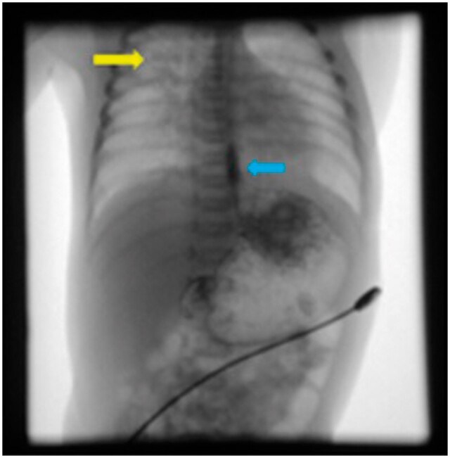
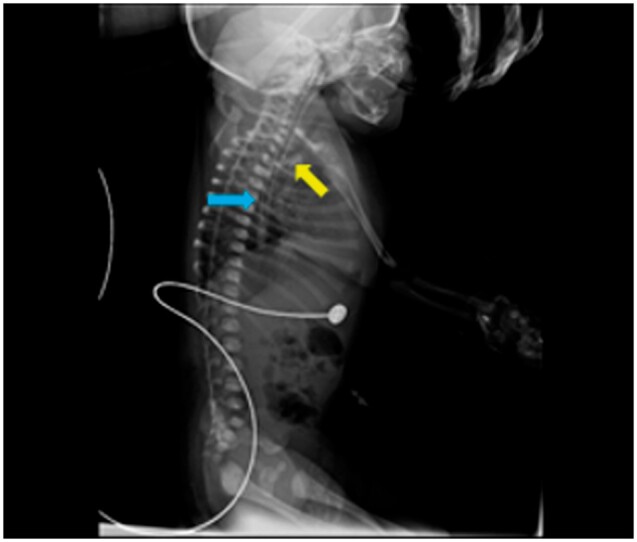
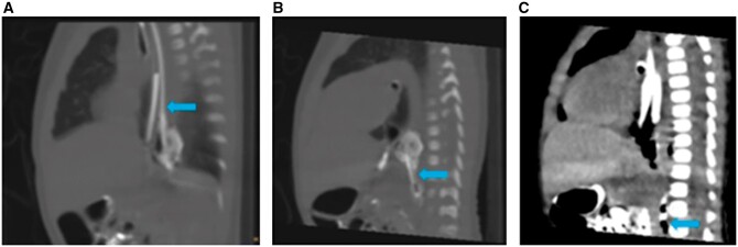
Intramural oesophageal dissection (IED) is an uncommon condition in newborns marked by the separation of the mucosal and submucosal layers of the oesophageal wall, both transversely and longitudinally, which may or may not involve perforation. A neonate presented at 26 h of life with poor respiratory effort and lethargy. She was intubated and was put on mechanical ventilation. Radiograph of the neonate suggested malpositioned endotracheal tube. The fluoroscopic dye-study indicated gastroesophageal oesophageal reflux disease and nothing significant. On limited CT contrast study of thorax, a tubular structure was seen running just parallel to the oesophagus extending from the T2 to the T9 levels. Possibilities of a oesophageal duplication/IED were considered. The neonate underwent an endoscopy and gastrostomy on day of life (DOL) 9. On follow up at 3 months a repeat limited CT study was done with instillation of water-soluble contrast. The previously seen tubular structure running parallel to the oesophagus was no longer seen. This finding suggested a healed IED. This case report emphasizes the significance of multimodality imaging in the diagnosis of this condition.

 求助内容:
求助内容: 应助结果提醒方式:


