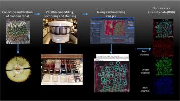
利用荧光显微镜和不同的传统及荧光染色方法分析结构组成的规程

IF 1.6 Q2 MULTIDISCIPLINARY SCIENCES
MethodsX Pub Date : 2024-10-10 DOI:10.1016/j.mex.2024.102999
Agustín Maceda , Agustina Rosa Andrés-Hernández , Teresa Terrazas
{"title":"利用荧光显微镜和不同的传统及荧光染色方法分析结构组成的规程","authors":"Agustín Maceda ,&nbsp;Agustina Rosa Andrés-Hernández ,&nbsp;Teresa Terrazas","doi":"10.1016/j.mex.2024.102999","DOIUrl":null,"url":null,"abstract":"<div><div>The protocol shows the effectiveness of using safranin-fast green stain for fluorescence microscopy. This staining technique has been used in conventional microscopy to perform anatomical characterizations of plants. However, this protocol describes the procedure for using samples stained with safranin-fast green in conjunction with fluorescence microscopy. The strength of the protocol lies in the fact that the samples are permanent and allows for effective differentiation of lignified and cellulosic walls unlike conventional fluorescence microscopy stains such as Congo red-acridine orange, calcofluor, and autofluorescence. The protocol for making fluorescence intensity measurements is also standardized, allowing the data to be used for statistical analysis and inference about the chemical composition of plant cell walls.</div></div>","PeriodicalId":18446,"journal":{"name":"MethodsX","volume":"13 ","pages":"Article 102999"},"PeriodicalIF":1.6000,"publicationDate":"2024-10-10","publicationTypes":"Journal Article","fieldsOfStudy":null,"isOpenAccess":false,"openAccessPdf":"","citationCount":"0","resultStr":"{\"title\":\"Protocol to analyse the structural composition by fluorescence microscopy and different conventional and fluorescence staining methods\",\"authors\":\"Agustín Maceda ,&nbsp;Agustina Rosa Andrés-Hernández ,&nbsp;Teresa Terrazas\",\"doi\":\"10.1016/j.mex.2024.102999\",\"DOIUrl\":null,\"url\":null,\"abstract\":\"<div><div>The protocol shows the effectiveness of using safranin-fast green stain for fluorescence microscopy. This staining technique has been used in conventional microscopy to perform anatomical characterizations of plants. However, this protocol describes the procedure for using samples stained with safranin-fast green in conjunction with fluorescence microscopy. The strength of the protocol lies in the fact that the samples are permanent and allows for effective differentiation of lignified and cellulosic walls unlike conventional fluorescence microscopy stains such as Congo red-acridine orange, calcofluor, and autofluorescence. The protocol for making fluorescence intensity measurements is also standardized, allowing the data to be used for statistical analysis and inference about the chemical composition of plant cell walls.</div></div>\",\"PeriodicalId\":18446,\"journal\":{\"name\":\"MethodsX\",\"volume\":\"13 \",\"pages\":\"Article 102999\"},\"PeriodicalIF\":1.6000,\"publicationDate\":\"2024-10-10\",\"publicationTypes\":\"Journal Article\",\"fieldsOfStudy\":null,\"isOpenAccess\":false,\"openAccessPdf\":\"\",\"citationCount\":\"0\",\"resultStr\":null,\"platform\":\"Semanticscholar\",\"paperid\":null,\"PeriodicalName\":\"MethodsX\",\"FirstCategoryId\":\"1085\",\"ListUrlMain\":\"https://www.sciencedirect.com/science/article/pii/S2215016124004503\",\"RegionNum\":0,\"RegionCategory\":null,\"ArticlePicture\":[],\"TitleCN\":null,\"AbstractTextCN\":null,\"PMCID\":null,\"EPubDate\":\"\",\"PubModel\":\"\",\"JCR\":\"Q2\",\"JCRName\":\"MULTIDISCIPLINARY SCIENCES\",\"Score\":null,\"Total\":0}","platform":"Semanticscholar","paperid":null,"PeriodicalName":"MethodsX","FirstCategoryId":"1085","ListUrlMain":"https://www.sciencedirect.com/science/article/pii/S2215016124004503","RegionNum":0,"RegionCategory":null,"ArticlePicture":[],"TitleCN":null,"AbstractTextCN":null,"PMCID":null,"EPubDate":"","PubModel":"","JCR":"Q2","JCRName":"MULTIDISCIPLINARY SCIENCES","Score":null,"Total":0}
引用次数: 0

摘要

该方案显示了在荧光显微镜下使用黄绿素-快绿染色剂的有效性。这种染色技术已在传统显微镜中用于植物的解剖特征描述。不过,本方案介绍的是将使用快绿素染色的样本与荧光显微镜结合使用的程序。与刚果红-吖啶橙、钙荧光和自发荧光等传统荧光显微染色法不同,本方案的优势在于样品是永久性的,可有效区分木质化壁和纤维素壁。荧光强度测量的规程也是标准化的,可将数据用于统计分析和推断植物细胞壁的化学成分。
本文章由计算机程序翻译,如有差异,请以英文原文为准。

Protocol to analyse the structural composition by fluorescence microscopy and different conventional and fluorescence staining methods

Protocol to analyse the structural composition by fluorescence microscopy and different conventional and fluorescence staining methods
The protocol shows the effectiveness of using safranin-fast green stain for fluorescence microscopy. This staining technique has been used in conventional microscopy to perform anatomical characterizations of plants. However, this protocol describes the procedure for using samples stained with safranin-fast green in conjunction with fluorescence microscopy. The strength of the protocol lies in the fact that the samples are permanent and allows for effective differentiation of lignified and cellulosic walls unlike conventional fluorescence microscopy stains such as Congo red-acridine orange, calcofluor, and autofluorescence. The protocol for making fluorescence intensity measurements is also standardized, allowing the data to be used for statistical analysis and inference about the chemical composition of plant cell walls.
求助全文
通过发布文献求助,成功后即可免费获取论文全文。 去求助
来源期刊
MethodsX
MethodsX Health Professions-Medical Laboratory Technology
CiteScore
3.60
自引率
5.30%
发文量
314
审稿时长
7 weeks
期刊介绍:
×
引用
GB/T 7714-2015
复制
MLA
复制
APA
复制
导出至
BibTeX EndNote RefMan NoteFirst NoteExpress
×
提示
您的信息不完整,为了账户安全,请先补充。
现在去补充
×
提示
您因"违规操作"
具体请查看互助需知
我知道了
×
提示
确定
请完成安全验证×
copy
已复制链接
快去分享给好友吧!
我知道了
右上角分享
点击右上角分享
0
联系我们:info@booksci.cn Book学术提供免费学术资源搜索服务,方便国内外学者检索中英文文献。致力于提供最便捷和优质的服务体验。 Copyright © 2023 布克学术 All rights reserved.
京ICP备2023020795号-1
ghs 京公网安备 11010802042870号
Book学术文献互助
Book学术文献互助群
群 号:481959085
Book学术官方微信