João D. Nunes , Diana Montezuma , Domingos Oliveira , Tania Pereira , Jaime S. Cardoso
{"title":"细胞核实例分割和分类调查:利用上下文和注意力","authors":"João D. Nunes , Diana Montezuma , Domingos Oliveira , Tania Pereira , Jaime S. Cardoso","doi":"10.1016/j.media.2024.103360","DOIUrl":null,"url":null,"abstract":"<div><div>Nuclear-derived morphological features and biomarkers provide relevant insights regarding the tumour microenvironment, while also allowing diagnosis and prognosis in specific cancer types. However, manually annotating nuclei from the gigapixel Haematoxylin and Eosin (H&E)-stained Whole Slide Images (WSIs) is a laborious and costly task, meaning automated algorithms for cell nuclei instance segmentation and classification could alleviate the workload of pathologists and clinical researchers and at the same time facilitate the automatic extraction of clinically interpretable features for artificial intelligence (AI) tools. But due to high intra- and inter-class variability of nuclei morphological and chromatic features, as well as H&E-stains susceptibility to artefacts, state-of-the-art algorithms cannot correctly detect and classify instances with the necessary performance. In this work, we hypothesize context and attention inductive biases in artificial neural networks (ANNs) could increase the performance and generalization of algorithms for cell nuclei instance segmentation and classification. To understand the advantages, use-cases, and limitations of context and attention-based mechanisms in instance segmentation and classification, we start by reviewing works in computer vision and medical imaging. We then conduct a thorough survey on context and attention methods for cell nuclei instance segmentation and classification from H&E-stained microscopy imaging, while providing a comprehensive discussion of the challenges being tackled with context and attention. Besides, we illustrate some limitations of current approaches and present ideas for future research. As a case study, we extend both a general (Mask-RCNN) and a customized (HoVer-Net) instance segmentation and classification methods with context- and attention-based mechanisms and perform a comparative analysis on a multicentre dataset for colon nuclei identification and counting.</div><div>Although pathologists rely on context at multiple levels while paying attention to specific Regions of Interest (RoIs) when analysing and annotating WSIs, our findings suggest translating that domain knowledge into algorithm design is no trivial task, but to fully exploit these mechanisms in ANNs, the scientific understanding of these methods should first be addressed.</div></div>","PeriodicalId":18328,"journal":{"name":"Medical image analysis","volume":"99 ","pages":"Article 103360"},"PeriodicalIF":10.7000,"publicationDate":"2024-10-05","publicationTypes":"Journal Article","fieldsOfStudy":null,"isOpenAccess":false,"openAccessPdf":"","citationCount":"0","resultStr":"{\"title\":\"A survey on cell nuclei instance segmentation and classification: Leveraging context and attention\",\"authors\":\"João D. Nunes , Diana Montezuma , Domingos Oliveira , Tania Pereira , Jaime S. Cardoso\",\"doi\":\"10.1016/j.media.2024.103360\",\"DOIUrl\":null,\"url\":null,\"abstract\":\"<div><div>Nuclear-derived morphological features and biomarkers provide relevant insights regarding the tumour microenvironment, while also allowing diagnosis and prognosis in specific cancer types. However, manually annotating nuclei from the gigapixel Haematoxylin and Eosin (H&E)-stained Whole Slide Images (WSIs) is a laborious and costly task, meaning automated algorithms for cell nuclei instance segmentation and classification could alleviate the workload of pathologists and clinical researchers and at the same time facilitate the automatic extraction of clinically interpretable features for artificial intelligence (AI) tools. But due to high intra- and inter-class variability of nuclei morphological and chromatic features, as well as H&E-stains susceptibility to artefacts, state-of-the-art algorithms cannot correctly detect and classify instances with the necessary performance. In this work, we hypothesize context and attention inductive biases in artificial neural networks (ANNs) could increase the performance and generalization of algorithms for cell nuclei instance segmentation and classification. To understand the advantages, use-cases, and limitations of context and attention-based mechanisms in instance segmentation and classification, we start by reviewing works in computer vision and medical imaging. We then conduct a thorough survey on context and attention methods for cell nuclei instance segmentation and classification from H&E-stained microscopy imaging, while providing a comprehensive discussion of the challenges being tackled with context and attention. Besides, we illustrate some limitations of current approaches and present ideas for future research. As a case study, we extend both a general (Mask-RCNN) and a customized (HoVer-Net) instance segmentation and classification methods with context- and attention-based mechanisms and perform a comparative analysis on a multicentre dataset for colon nuclei identification and counting.</div><div>Although pathologists rely on context at multiple levels while paying attention to specific Regions of Interest (RoIs) when analysing and annotating WSIs, our findings suggest translating that domain knowledge into algorithm design is no trivial task, but to fully exploit these mechanisms in ANNs, the scientific understanding of these methods should first be addressed.</div></div>\",\"PeriodicalId\":18328,\"journal\":{\"name\":\"Medical image analysis\",\"volume\":\"99 \",\"pages\":\"Article 103360\"},\"PeriodicalIF\":10.7000,\"publicationDate\":\"2024-10-05\",\"publicationTypes\":\"Journal Article\",\"fieldsOfStudy\":null,\"isOpenAccess\":false,\"openAccessPdf\":\"\",\"citationCount\":\"0\",\"resultStr\":null,\"platform\":\"Semanticscholar\",\"paperid\":null,\"PeriodicalName\":\"Medical image analysis\",\"FirstCategoryId\":\"5\",\"ListUrlMain\":\"https://www.sciencedirect.com/science/article/pii/S1361841524002858\",\"RegionNum\":1,\"RegionCategory\":\"医学\",\"ArticlePicture\":[],\"TitleCN\":null,\"AbstractTextCN\":null,\"PMCID\":null,\"EPubDate\":\"\",\"PubModel\":\"\",\"JCR\":\"Q1\",\"JCRName\":\"COMPUTER SCIENCE, ARTIFICIAL INTELLIGENCE\",\"Score\":null,\"Total\":0}","platform":"Semanticscholar","paperid":null,"PeriodicalName":"Medical image analysis","FirstCategoryId":"5","ListUrlMain":"https://www.sciencedirect.com/science/article/pii/S1361841524002858","RegionNum":1,"RegionCategory":"医学","ArticlePicture":[],"TitleCN":null,"AbstractTextCN":null,"PMCID":null,"EPubDate":"","PubModel":"","JCR":"Q1","JCRName":"COMPUTER SCIENCE, ARTIFICIAL INTELLIGENCE","Score":null,"Total":0}
A survey on cell nuclei instance segmentation and classification: Leveraging context and attention
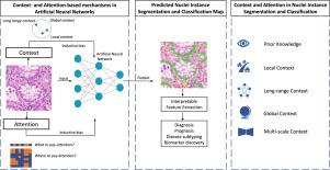
Nuclear-derived morphological features and biomarkers provide relevant insights regarding the tumour microenvironment, while also allowing diagnosis and prognosis in specific cancer types. However, manually annotating nuclei from the gigapixel Haematoxylin and Eosin (H&E)-stained Whole Slide Images (WSIs) is a laborious and costly task, meaning automated algorithms for cell nuclei instance segmentation and classification could alleviate the workload of pathologists and clinical researchers and at the same time facilitate the automatic extraction of clinically interpretable features for artificial intelligence (AI) tools. But due to high intra- and inter-class variability of nuclei morphological and chromatic features, as well as H&E-stains susceptibility to artefacts, state-of-the-art algorithms cannot correctly detect and classify instances with the necessary performance. In this work, we hypothesize context and attention inductive biases in artificial neural networks (ANNs) could increase the performance and generalization of algorithms for cell nuclei instance segmentation and classification. To understand the advantages, use-cases, and limitations of context and attention-based mechanisms in instance segmentation and classification, we start by reviewing works in computer vision and medical imaging. We then conduct a thorough survey on context and attention methods for cell nuclei instance segmentation and classification from H&E-stained microscopy imaging, while providing a comprehensive discussion of the challenges being tackled with context and attention. Besides, we illustrate some limitations of current approaches and present ideas for future research. As a case study, we extend both a general (Mask-RCNN) and a customized (HoVer-Net) instance segmentation and classification methods with context- and attention-based mechanisms and perform a comparative analysis on a multicentre dataset for colon nuclei identification and counting.
Although pathologists rely on context at multiple levels while paying attention to specific Regions of Interest (RoIs) when analysing and annotating WSIs, our findings suggest translating that domain knowledge into algorithm design is no trivial task, but to fully exploit these mechanisms in ANNs, the scientific understanding of these methods should first be addressed.
期刊介绍:
Medical Image Analysis serves as a platform for sharing new research findings in the realm of medical and biological image analysis, with a focus on applications of computer vision, virtual reality, and robotics to biomedical imaging challenges. The journal prioritizes the publication of high-quality, original papers contributing to the fundamental science of processing, analyzing, and utilizing medical and biological images. It welcomes approaches utilizing biomedical image datasets across all spatial scales, from molecular/cellular imaging to tissue/organ imaging.