{"title":"影像学轴性脊柱关节炎中关节面结构病变的不同特征和发展模式。","authors":"Simin Liao, Liuquan Cheng, Zheng Zhao, Jian Zhu, Feng Huang","doi":"10.1177/1759720X241281201","DOIUrl":null,"url":null,"abstract":"<p><strong>Background: </strong>Both vertebral bodies and posterior elements of the vertebrae (facet joints, FJ) can engage in bone formation in radiographic axial spondyloarthritis (r-axSpA). However, little is known about the specific structural lesions and progression patterns of FJs in r-axSpA.</p><p><strong>Objectives: </strong>To identify specific lesions related to r-axSpA and to investigate the distinct progression patterns by comparing the FJ changes of r-axSpA with that of diffuse idiopathic skeletal hyperostosis (DISH), osteoarthritis (OA), and control group (CG).</p><p><strong>Design: </strong>Single-center, retrospective study. Longitudinal imaging data were retrieved and collected.</p><p><strong>Methods: </strong>Age- and sex-matched patients with complete thoracic and lumbar spine computed tomography (CT) data were included and their bilateral FJs were assessed. FJ changes were divided into erosions, ankylosis, joint-space narrowing, osteophytes, subchondral sclerosis, subchondral cysts, and vacuum phenomena. Average progressed year was defined as \"number of changed vertebrae × interval years\"/number of changed vertebrae.</p><p><strong>Results: </strong>In all, 50 patients in each group were included. Subchondral cysts and vacuum phenomena were not observed. Bilateral FJ ankylosis (FJA)/erosions in the thoracic and lumbar spine, and unilateral ankylosis/erosions in T1-4, T9-12 were significantly more common in r-axSpA. Joint-space narrowing/osteophytes/subchondral sclerosis were significantly more common in DISH and OA. FJ lesions progressed in 56.34% of vertebrae of r-axSpA. The most common pattern was \"FJ normal advanced to ankylosis\" (17.54%) which required 2.63 years. It was followed by \"erosions advanced to ankylosis\" (12.3%) which took 2.05 years, and by \"normal FJ advanced to erosions\" (11.04%) which took 2.29 years, respectively. Degenerative changes could also progress to FJ erosions/ankylosis (24.83%). The majority pattern in DISH/OA was \"FJ changes advanced to subchondral sclerosis/osteophytes/joint-space narrowing.\"</p><p><strong>Conclusion: </strong>Bilateral FJA/erosions are r-axSpA-specific lesions. The specific progression pattern for r-axSpA was \"FJ changes advanced to ankylosis/erosions.\" Repeated CT examination in intervals of at least 2 years will be more appropriate for monitoring FJ progression.</p>","PeriodicalId":23056,"journal":{"name":"Therapeutic Advances in Musculoskeletal Disease","volume":"16 ","pages":"1759720X241281201"},"PeriodicalIF":4.1000,"publicationDate":"2024-09-28","publicationTypes":"Journal Article","fieldsOfStudy":null,"isOpenAccess":false,"openAccessPdf":"https://www.ncbi.nlm.nih.gov/pmc/articles/PMC11450570/pdf/","citationCount":"0","resultStr":"{\"title\":\"Distinct characteristics and progression patterns of facet joint structural lesions in radiographic axial spondyloarthritis.\",\"authors\":\"Simin Liao, Liuquan Cheng, Zheng Zhao, Jian Zhu, Feng Huang\",\"doi\":\"10.1177/1759720X241281201\",\"DOIUrl\":null,\"url\":null,\"abstract\":\"<p><strong>Background: </strong>Both vertebral bodies and posterior elements of the vertebrae (facet joints, FJ) can engage in bone formation in radiographic axial spondyloarthritis (r-axSpA). However, little is known about the specific structural lesions and progression patterns of FJs in r-axSpA.</p><p><strong>Objectives: </strong>To identify specific lesions related to r-axSpA and to investigate the distinct progression patterns by comparing the FJ changes of r-axSpA with that of diffuse idiopathic skeletal hyperostosis (DISH), osteoarthritis (OA), and control group (CG).</p><p><strong>Design: </strong>Single-center, retrospective study. Longitudinal imaging data were retrieved and collected.</p><p><strong>Methods: </strong>Age- and sex-matched patients with complete thoracic and lumbar spine computed tomography (CT) data were included and their bilateral FJs were assessed. FJ changes were divided into erosions, ankylosis, joint-space narrowing, osteophytes, subchondral sclerosis, subchondral cysts, and vacuum phenomena. Average progressed year was defined as \\\"number of changed vertebrae × interval years\\\"/number of changed vertebrae.</p><p><strong>Results: </strong>In all, 50 patients in each group were included. Subchondral cysts and vacuum phenomena were not observed. Bilateral FJ ankylosis (FJA)/erosions in the thoracic and lumbar spine, and unilateral ankylosis/erosions in T1-4, T9-12 were significantly more common in r-axSpA. Joint-space narrowing/osteophytes/subchondral sclerosis were significantly more common in DISH and OA. FJ lesions progressed in 56.34% of vertebrae of r-axSpA. The most common pattern was \\\"FJ normal advanced to ankylosis\\\" (17.54%) which required 2.63 years. It was followed by \\\"erosions advanced to ankylosis\\\" (12.3%) which took 2.05 years, and by \\\"normal FJ advanced to erosions\\\" (11.04%) which took 2.29 years, respectively. Degenerative changes could also progress to FJ erosions/ankylosis (24.83%). The majority pattern in DISH/OA was \\\"FJ changes advanced to subchondral sclerosis/osteophytes/joint-space narrowing.\\\"</p><p><strong>Conclusion: </strong>Bilateral FJA/erosions are r-axSpA-specific lesions. The specific progression pattern for r-axSpA was \\\"FJ changes advanced to ankylosis/erosions.\\\" Repeated CT examination in intervals of at least 2 years will be more appropriate for monitoring FJ progression.</p>\",\"PeriodicalId\":23056,\"journal\":{\"name\":\"Therapeutic Advances in Musculoskeletal Disease\",\"volume\":\"16 \",\"pages\":\"1759720X241281201\"},\"PeriodicalIF\":4.1000,\"publicationDate\":\"2024-09-28\",\"publicationTypes\":\"Journal Article\",\"fieldsOfStudy\":null,\"isOpenAccess\":false,\"openAccessPdf\":\"https://www.ncbi.nlm.nih.gov/pmc/articles/PMC11450570/pdf/\",\"citationCount\":\"0\",\"resultStr\":null,\"platform\":\"Semanticscholar\",\"paperid\":null,\"PeriodicalName\":\"Therapeutic Advances in Musculoskeletal Disease\",\"FirstCategoryId\":\"3\",\"ListUrlMain\":\"https://doi.org/10.1177/1759720X241281201\",\"RegionNum\":2,\"RegionCategory\":\"医学\",\"ArticlePicture\":[],\"TitleCN\":null,\"AbstractTextCN\":null,\"PMCID\":null,\"EPubDate\":\"2024/1/1 0:00:00\",\"PubModel\":\"eCollection\",\"JCR\":\"Q2\",\"JCRName\":\"RHEUMATOLOGY\",\"Score\":null,\"Total\":0}","platform":"Semanticscholar","paperid":null,"PeriodicalName":"Therapeutic Advances in Musculoskeletal Disease","FirstCategoryId":"3","ListUrlMain":"https://doi.org/10.1177/1759720X241281201","RegionNum":2,"RegionCategory":"医学","ArticlePicture":[],"TitleCN":null,"AbstractTextCN":null,"PMCID":null,"EPubDate":"2024/1/1 0:00:00","PubModel":"eCollection","JCR":"Q2","JCRName":"RHEUMATOLOGY","Score":null,"Total":0}
Distinct characteristics and progression patterns of facet joint structural lesions in radiographic axial spondyloarthritis.
Background: Both vertebral bodies and posterior elements of the vertebrae (facet joints, FJ) can engage in bone formation in radiographic axial spondyloarthritis (r-axSpA). However, little is known about the specific structural lesions and progression patterns of FJs in r-axSpA.
Objectives: To identify specific lesions related to r-axSpA and to investigate the distinct progression patterns by comparing the FJ changes of r-axSpA with that of diffuse idiopathic skeletal hyperostosis (DISH), osteoarthritis (OA), and control group (CG).
Design: Single-center, retrospective study. Longitudinal imaging data were retrieved and collected.
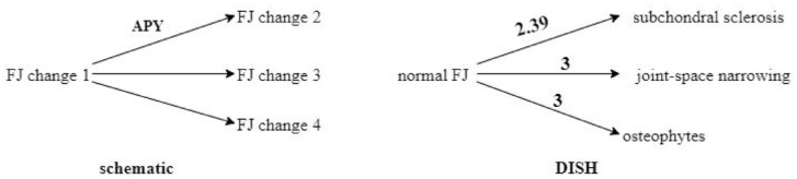
Methods: Age- and sex-matched patients with complete thoracic and lumbar spine computed tomography (CT) data were included and their bilateral FJs were assessed. FJ changes were divided into erosions, ankylosis, joint-space narrowing, osteophytes, subchondral sclerosis, subchondral cysts, and vacuum phenomena. Average progressed year was defined as "number of changed vertebrae × interval years"/number of changed vertebrae.
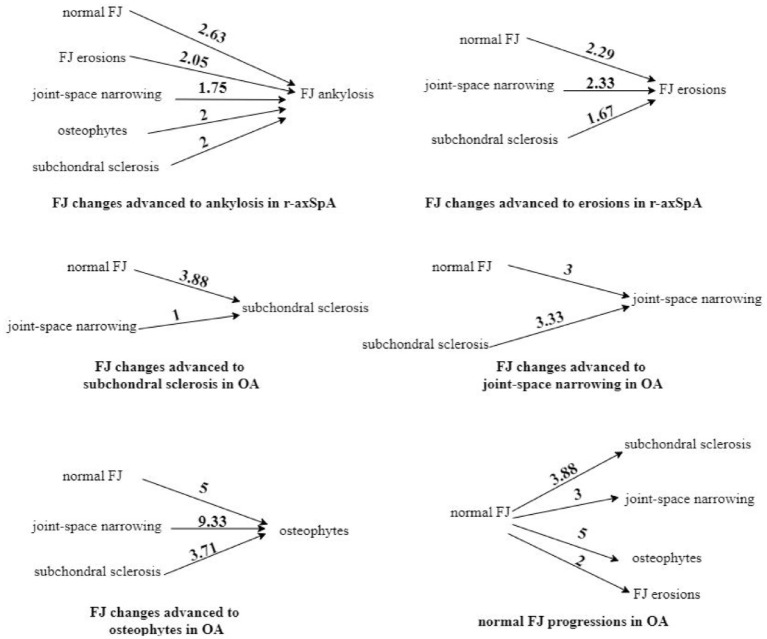
Results: In all, 50 patients in each group were included. Subchondral cysts and vacuum phenomena were not observed. Bilateral FJ ankylosis (FJA)/erosions in the thoracic and lumbar spine, and unilateral ankylosis/erosions in T1-4, T9-12 were significantly more common in r-axSpA. Joint-space narrowing/osteophytes/subchondral sclerosis were significantly more common in DISH and OA. FJ lesions progressed in 56.34% of vertebrae of r-axSpA. The most common pattern was "FJ normal advanced to ankylosis" (17.54%) which required 2.63 years. It was followed by "erosions advanced to ankylosis" (12.3%) which took 2.05 years, and by "normal FJ advanced to erosions" (11.04%) which took 2.29 years, respectively. Degenerative changes could also progress to FJ erosions/ankylosis (24.83%). The majority pattern in DISH/OA was "FJ changes advanced to subchondral sclerosis/osteophytes/joint-space narrowing."
Conclusion: Bilateral FJA/erosions are r-axSpA-specific lesions. The specific progression pattern for r-axSpA was "FJ changes advanced to ankylosis/erosions." Repeated CT examination in intervals of at least 2 years will be more appropriate for monitoring FJ progression.
期刊介绍:
Therapeutic Advances in Musculoskeletal Disease delivers the highest quality peer-reviewed articles, reviews, and scholarly comment on pioneering efforts and innovative studies across all areas of musculoskeletal disease.
 求助内容:
求助内容: 应助结果提醒方式:


