Michael Chen, Krit Suwannaphoom, Yas Sanaiha, Yuan Luo, Peyman Benharash, Michael C. Fishbein, René R. Sevag Packard
{"title":"电化学阻抗谱分析揭示了人类冠状动脉疾病的高风险动脉粥样硬化特征。","authors":"Michael Chen, Krit Suwannaphoom, Yas Sanaiha, Yuan Luo, Peyman Benharash, Michael C. Fishbein, René R. Sevag Packard","doi":"10.1096/fj.202401200R","DOIUrl":null,"url":null,"abstract":"<p>Coronary plaque rupture remains the prominent mechanism of myocardial infarction. Accurate identification of rupture-prone plaque may improve clinical management. This study assessed the discriminatory performance of electrochemical impedance spectroscopy (EIS) in human cardiac explants to detect high-risk atherosclerotic features that portend rupture risk. In this single-center, prospective study, <i>n</i> = 26 cardiac explants were collected for EIS interrogation of the three major coronary arteries. Vessels in which advancement of the EIS catheter without iatrogenic plaque disruption was rendered impossible were not assessed. <i>N</i> = 61 vessels underwent EIS measurement and histological analyses. Plaques were dichotomized according to previously established high rupture-risk parameter thresholds. Diagnostic performance was determined via receiver operating characteristic areas-under-the-curve (AUC). Necrotic cores were identified in <i>n</i> = 19 vessels (median area 1.53 mm<sup>2</sup>) with a median fibrous cap thickness of 62 μm. Impedance was significantly greater in plaques with necrotic core area ≥1.75 mm<sup>2</sup> versus <1.75 mm<sup>2</sup> (19.8 ± 4.4 kΩ vs. 7.2 ± 1.0 kΩ, <i>p</i> = .019), fibrous cap thickness ≤65 μm versus >65 μm (19.1 ± 3.5 kΩ vs. 6.5 ± 0.9 kΩ, <i>p</i> = .004), and ≥20 macrophages per 0.3 mm-diameter high-power field (HPF) versus <20 macrophages per HPF (19.8 ± 4.1 kΩ vs. 10.2 ± 0.9 kΩ, <i>p</i> = .002). Impedance identified necrotic core area ≥1.75 mm<sup>2</sup>, fibrous cap thickness ≤65 μm, and ≥20 macrophages per HPF with AUCs of 0.889 (95% CI: 0.716–1.000) (<i>p</i> = .013), 0.852 (0.646–1.000) (<i>p</i> = .025), and 0.835 (0.577–1.000) (<i>p</i> = .028), respectively. Further, phase delay discriminated severe stenosis (≥70%) with an AUC of 0.767 (0.573–0.962) (<i>p</i> = .035). EIS discriminates high-risk atherosclerotic features that portend plaque rupture in human coronary artery disease and may serve as a complementary modality for angiography-guided atherosclerosis evaluation.</p>","PeriodicalId":50455,"journal":{"name":"The FASEB Journal","volume":"38 18","pages":""},"PeriodicalIF":4.4000,"publicationDate":"2024-09-24","publicationTypes":"Journal Article","fieldsOfStudy":null,"isOpenAccess":false,"openAccessPdf":"","citationCount":"0","resultStr":"{\"title\":\"Electrochemical impedance spectroscopy unmasks high-risk atherosclerotic features in human coronary artery disease\",\"authors\":\"Michael Chen, Krit Suwannaphoom, Yas Sanaiha, Yuan Luo, Peyman Benharash, Michael C. Fishbein, René R. Sevag Packard\",\"doi\":\"10.1096/fj.202401200R\",\"DOIUrl\":null,\"url\":null,\"abstract\":\"<p>Coronary plaque rupture remains the prominent mechanism of myocardial infarction. Accurate identification of rupture-prone plaque may improve clinical management. This study assessed the discriminatory performance of electrochemical impedance spectroscopy (EIS) in human cardiac explants to detect high-risk atherosclerotic features that portend rupture risk. In this single-center, prospective study, <i>n</i> = 26 cardiac explants were collected for EIS interrogation of the three major coronary arteries. Vessels in which advancement of the EIS catheter without iatrogenic plaque disruption was rendered impossible were not assessed. <i>N</i> = 61 vessels underwent EIS measurement and histological analyses. Plaques were dichotomized according to previously established high rupture-risk parameter thresholds. Diagnostic performance was determined via receiver operating characteristic areas-under-the-curve (AUC). Necrotic cores were identified in <i>n</i> = 19 vessels (median area 1.53 mm<sup>2</sup>) with a median fibrous cap thickness of 62 μm. Impedance was significantly greater in plaques with necrotic core area ≥1.75 mm<sup>2</sup> versus <1.75 mm<sup>2</sup> (19.8 ± 4.4 kΩ vs. 7.2 ± 1.0 kΩ, <i>p</i> = .019), fibrous cap thickness ≤65 μm versus >65 μm (19.1 ± 3.5 kΩ vs. 6.5 ± 0.9 kΩ, <i>p</i> = .004), and ≥20 macrophages per 0.3 mm-diameter high-power field (HPF) versus <20 macrophages per HPF (19.8 ± 4.1 kΩ vs. 10.2 ± 0.9 kΩ, <i>p</i> = .002). Impedance identified necrotic core area ≥1.75 mm<sup>2</sup>, fibrous cap thickness ≤65 μm, and ≥20 macrophages per HPF with AUCs of 0.889 (95% CI: 0.716–1.000) (<i>p</i> = .013), 0.852 (0.646–1.000) (<i>p</i> = .025), and 0.835 (0.577–1.000) (<i>p</i> = .028), respectively. Further, phase delay discriminated severe stenosis (≥70%) with an AUC of 0.767 (0.573–0.962) (<i>p</i> = .035). EIS discriminates high-risk atherosclerotic features that portend plaque rupture in human coronary artery disease and may serve as a complementary modality for angiography-guided atherosclerosis evaluation.</p>\",\"PeriodicalId\":50455,\"journal\":{\"name\":\"The FASEB Journal\",\"volume\":\"38 18\",\"pages\":\"\"},\"PeriodicalIF\":4.4000,\"publicationDate\":\"2024-09-24\",\"publicationTypes\":\"Journal Article\",\"fieldsOfStudy\":null,\"isOpenAccess\":false,\"openAccessPdf\":\"\",\"citationCount\":\"0\",\"resultStr\":null,\"platform\":\"Semanticscholar\",\"paperid\":null,\"PeriodicalName\":\"The FASEB Journal\",\"FirstCategoryId\":\"99\",\"ListUrlMain\":\"https://onlinelibrary.wiley.com/doi/10.1096/fj.202401200R\",\"RegionNum\":2,\"RegionCategory\":\"生物学\",\"ArticlePicture\":[],\"TitleCN\":null,\"AbstractTextCN\":null,\"PMCID\":null,\"EPubDate\":\"\",\"PubModel\":\"\",\"JCR\":\"Q2\",\"JCRName\":\"BIOCHEMISTRY & MOLECULAR BIOLOGY\",\"Score\":null,\"Total\":0}","platform":"Semanticscholar","paperid":null,"PeriodicalName":"The FASEB Journal","FirstCategoryId":"99","ListUrlMain":"https://onlinelibrary.wiley.com/doi/10.1096/fj.202401200R","RegionNum":2,"RegionCategory":"生物学","ArticlePicture":[],"TitleCN":null,"AbstractTextCN":null,"PMCID":null,"EPubDate":"","PubModel":"","JCR":"Q2","JCRName":"BIOCHEMISTRY & MOLECULAR BIOLOGY","Score":null,"Total":0}
Electrochemical impedance spectroscopy unmasks high-risk atherosclerotic features in human coronary artery disease
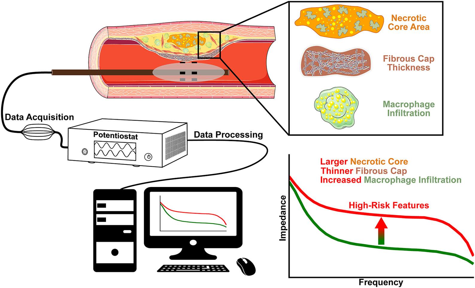
Coronary plaque rupture remains the prominent mechanism of myocardial infarction. Accurate identification of rupture-prone plaque may improve clinical management. This study assessed the discriminatory performance of electrochemical impedance spectroscopy (EIS) in human cardiac explants to detect high-risk atherosclerotic features that portend rupture risk. In this single-center, prospective study, n = 26 cardiac explants were collected for EIS interrogation of the three major coronary arteries. Vessels in which advancement of the EIS catheter without iatrogenic plaque disruption was rendered impossible were not assessed. N = 61 vessels underwent EIS measurement and histological analyses. Plaques were dichotomized according to previously established high rupture-risk parameter thresholds. Diagnostic performance was determined via receiver operating characteristic areas-under-the-curve (AUC). Necrotic cores were identified in n = 19 vessels (median area 1.53 mm2) with a median fibrous cap thickness of 62 μm. Impedance was significantly greater in plaques with necrotic core area ≥1.75 mm2 versus <1.75 mm2 (19.8 ± 4.4 kΩ vs. 7.2 ± 1.0 kΩ, p = .019), fibrous cap thickness ≤65 μm versus >65 μm (19.1 ± 3.5 kΩ vs. 6.5 ± 0.9 kΩ, p = .004), and ≥20 macrophages per 0.3 mm-diameter high-power field (HPF) versus <20 macrophages per HPF (19.8 ± 4.1 kΩ vs. 10.2 ± 0.9 kΩ, p = .002). Impedance identified necrotic core area ≥1.75 mm2, fibrous cap thickness ≤65 μm, and ≥20 macrophages per HPF with AUCs of 0.889 (95% CI: 0.716–1.000) (p = .013), 0.852 (0.646–1.000) (p = .025), and 0.835 (0.577–1.000) (p = .028), respectively. Further, phase delay discriminated severe stenosis (≥70%) with an AUC of 0.767 (0.573–0.962) (p = .035). EIS discriminates high-risk atherosclerotic features that portend plaque rupture in human coronary artery disease and may serve as a complementary modality for angiography-guided atherosclerosis evaluation.
期刊介绍:
The FASEB Journal publishes international, transdisciplinary research covering all fields of biology at every level of organization: atomic, molecular, cell, tissue, organ, organismic and population. While the journal strives to include research that cuts across the biological sciences, it also considers submissions that lie within one field, but may have implications for other fields as well. The journal seeks to publish basic and translational research, but also welcomes reports of pre-clinical and early clinical research. In addition to research, review, and hypothesis submissions, The FASEB Journal also seeks perspectives, commentaries, book reviews, and similar content related to the life sciences in its Up Front section.
 求助内容:
求助内容: 应助结果提醒方式:


