Alessandra Borgheresi, Elisa Cesari, Andrea Agostini, Myriam Badaloni, Sofia Balducci, Elisabetta Tola, Valeria Consoli, Andrea Palucci, Luca Burroni, Marina Carotti, Andrea Giovagnoni
{"title":"肺气肿:用双能量 CT 和肺部闪烁扫描评估肺部灌注情况","authors":"Alessandra Borgheresi, Elisa Cesari, Andrea Agostini, Myriam Badaloni, Sofia Balducci, Elisabetta Tola, Valeria Consoli, Andrea Palucci, Luca Burroni, Marina Carotti, Andrea Giovagnoni","doi":"10.1007/s11547-024-01883-y","DOIUrl":null,"url":null,"abstract":"<h3 data-test=\"abstract-sub-heading\">Aim</h3><p>To assess the correlation of quantitative data of pulmonary Perfused Blood Volume (PBV) on Dual-Energy CT (DECT) datasets in patients with moderate – severe Pulmonary Emphysema (PE) with Lung Perfusion Scintigraphy (LPS) as the reference standard. The secondary endpoints are the correlation between the CT densitometric analysis and the visual assessment of parenchymal destruction with PBV.</p><h3 data-test=\"abstract-sub-heading\">Materials and Methods</h3><p>Patients with moderate – severe PE candidate to Lung Volumetric Reduction (LVR), with available a pre-procedural LS and a contrast-enhanced DECT were retrospectively included. DECT studies were performed with a 3rd generation Dual-Source CT and the PBV was obtained with a 3-material decomposition algorithm. The CT densitometric analysis was performed with a dedicated commercial software (Pulmo3D). The Goddard Score was used for visual assessment. The perfusion LS were performed after the administration of albumin macroaggregates labeled with <sup>99m</sup>Technetium. The image revision was performed by two radiologists or nuclear medicine physicians blinded, respectively, to LS and DECT data. The statistical analysis was performed with nonparametric tests.</p><h3 data-test=\"abstract-sub-heading\">Results</h3><p>Thirty-one patients (18 males, median age 69 y.o., interquartile range 62–71 y.o.) with moderate – severe PE (Median Goddard Score 14/20 and 31% of emphysematous parenchyma at quantitative CT) candidate to LVR were retrospectively included. The median enhancement on PBV was 17 HU. Significant correlation coefficients were demonstrated between lung PBV and LS, poor in apical regions (Rho = 0.1–0.2) and fair (Rho = 0.3–0.5) in middle and lower regions. No significant correlations were recorded between the CT densitometric analysis, the visual score, and the PBV.</p><h3 data-test=\"abstract-sub-heading\">Conclusions</h3><p>Lung perfusion with PBV on DECT is feasible in patients with moderate – severe PE candidate to LVR, and has a poor to fair agreement with LPS.</p>","PeriodicalId":501689,"journal":{"name":"La radiologia medica","volume":"47 1","pages":""},"PeriodicalIF":0.0000,"publicationDate":"2024-09-10","publicationTypes":"Journal Article","fieldsOfStudy":null,"isOpenAccess":false,"openAccessPdf":"","citationCount":"0","resultStr":"{\"title\":\"Pulmonary emphysema: the assessment of lung perfusion with Dual-Energy CT and pulmonary scintigraphy\",\"authors\":\"Alessandra Borgheresi, Elisa Cesari, Andrea Agostini, Myriam Badaloni, Sofia Balducci, Elisabetta Tola, Valeria Consoli, Andrea Palucci, Luca Burroni, Marina Carotti, Andrea Giovagnoni\",\"doi\":\"10.1007/s11547-024-01883-y\",\"DOIUrl\":null,\"url\":null,\"abstract\":\"<h3 data-test=\\\"abstract-sub-heading\\\">Aim</h3><p>To assess the correlation of quantitative data of pulmonary Perfused Blood Volume (PBV) on Dual-Energy CT (DECT) datasets in patients with moderate – severe Pulmonary Emphysema (PE) with Lung Perfusion Scintigraphy (LPS) as the reference standard. The secondary endpoints are the correlation between the CT densitometric analysis and the visual assessment of parenchymal destruction with PBV.</p><h3 data-test=\\\"abstract-sub-heading\\\">Materials and Methods</h3><p>Patients with moderate – severe PE candidate to Lung Volumetric Reduction (LVR), with available a pre-procedural LS and a contrast-enhanced DECT were retrospectively included. DECT studies were performed with a 3rd generation Dual-Source CT and the PBV was obtained with a 3-material decomposition algorithm. The CT densitometric analysis was performed with a dedicated commercial software (Pulmo3D). The Goddard Score was used for visual assessment. The perfusion LS were performed after the administration of albumin macroaggregates labeled with <sup>99m</sup>Technetium. The image revision was performed by two radiologists or nuclear medicine physicians blinded, respectively, to LS and DECT data. The statistical analysis was performed with nonparametric tests.</p><h3 data-test=\\\"abstract-sub-heading\\\">Results</h3><p>Thirty-one patients (18 males, median age 69 y.o., interquartile range 62–71 y.o.) with moderate – severe PE (Median Goddard Score 14/20 and 31% of emphysematous parenchyma at quantitative CT) candidate to LVR were retrospectively included. The median enhancement on PBV was 17 HU. Significant correlation coefficients were demonstrated between lung PBV and LS, poor in apical regions (Rho = 0.1–0.2) and fair (Rho = 0.3–0.5) in middle and lower regions. No significant correlations were recorded between the CT densitometric analysis, the visual score, and the PBV.</p><h3 data-test=\\\"abstract-sub-heading\\\">Conclusions</h3><p>Lung perfusion with PBV on DECT is feasible in patients with moderate – severe PE candidate to LVR, and has a poor to fair agreement with LPS.</p>\",\"PeriodicalId\":501689,\"journal\":{\"name\":\"La radiologia medica\",\"volume\":\"47 1\",\"pages\":\"\"},\"PeriodicalIF\":0.0000,\"publicationDate\":\"2024-09-10\",\"publicationTypes\":\"Journal Article\",\"fieldsOfStudy\":null,\"isOpenAccess\":false,\"openAccessPdf\":\"\",\"citationCount\":\"0\",\"resultStr\":null,\"platform\":\"Semanticscholar\",\"paperid\":null,\"PeriodicalName\":\"La radiologia medica\",\"FirstCategoryId\":\"1085\",\"ListUrlMain\":\"https://doi.org/10.1007/s11547-024-01883-y\",\"RegionNum\":0,\"RegionCategory\":null,\"ArticlePicture\":[],\"TitleCN\":null,\"AbstractTextCN\":null,\"PMCID\":null,\"EPubDate\":\"\",\"PubModel\":\"\",\"JCR\":\"\",\"JCRName\":\"\",\"Score\":null,\"Total\":0}","platform":"Semanticscholar","paperid":null,"PeriodicalName":"La radiologia medica","FirstCategoryId":"1085","ListUrlMain":"https://doi.org/10.1007/s11547-024-01883-y","RegionNum":0,"RegionCategory":null,"ArticlePicture":[],"TitleCN":null,"AbstractTextCN":null,"PMCID":null,"EPubDate":"","PubModel":"","JCR":"","JCRName":"","Score":null,"Total":0}
Pulmonary emphysema: the assessment of lung perfusion with Dual-Energy CT and pulmonary scintigraphy
Aim
To assess the correlation of quantitative data of pulmonary Perfused Blood Volume (PBV) on Dual-Energy CT (DECT) datasets in patients with moderate – severe Pulmonary Emphysema (PE) with Lung Perfusion Scintigraphy (LPS) as the reference standard. The secondary endpoints are the correlation between the CT densitometric analysis and the visual assessment of parenchymal destruction with PBV.
Materials and Methods
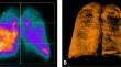
Patients with moderate – severe PE candidate to Lung Volumetric Reduction (LVR), with available a pre-procedural LS and a contrast-enhanced DECT were retrospectively included. DECT studies were performed with a 3rd generation Dual-Source CT and the PBV was obtained with a 3-material decomposition algorithm. The CT densitometric analysis was performed with a dedicated commercial software (Pulmo3D). The Goddard Score was used for visual assessment. The perfusion LS were performed after the administration of albumin macroaggregates labeled with 99mTechnetium. The image revision was performed by two radiologists or nuclear medicine physicians blinded, respectively, to LS and DECT data. The statistical analysis was performed with nonparametric tests.
Results
Thirty-one patients (18 males, median age 69 y.o., interquartile range 62–71 y.o.) with moderate – severe PE (Median Goddard Score 14/20 and 31% of emphysematous parenchyma at quantitative CT) candidate to LVR were retrospectively included. The median enhancement on PBV was 17 HU. Significant correlation coefficients were demonstrated between lung PBV and LS, poor in apical regions (Rho = 0.1–0.2) and fair (Rho = 0.3–0.5) in middle and lower regions. No significant correlations were recorded between the CT densitometric analysis, the visual score, and the PBV.
Conclusions
Lung perfusion with PBV on DECT is feasible in patients with moderate – severe PE candidate to LVR, and has a poor to fair agreement with LPS.

 求助内容:
求助内容: 应助结果提醒方式:


