Lorraine J. Lauwerends, Bo E. Zweedijk, Hidde A. Galema, Lisanne K. A. Neijenhuis, Neeltje G. Dekker-Ensink, Robert J. Baatenburg de Jong, Cornelis Verhoef, Shadhvi S. Bhairosingh, Peter J. K. Kuppen, Alexander L. Vahrmeijer, Tessa M. van Ginhoven, Senada Koljenović, Sjors A. Koppes, Denise E. Hilling, Stijn Keereweer
{"title":"头颈部恶性肿瘤中的肿瘤标记物表达,以确定术中分子近红外成像的潜在目标","authors":"Lorraine J. Lauwerends, Bo E. Zweedijk, Hidde A. Galema, Lisanne K. A. Neijenhuis, Neeltje G. Dekker-Ensink, Robert J. Baatenburg de Jong, Cornelis Verhoef, Shadhvi S. Bhairosingh, Peter J. K. Kuppen, Alexander L. Vahrmeijer, Tessa M. van Ginhoven, Senada Koljenović, Sjors A. Koppes, Denise E. Hilling, Stijn Keereweer","doi":"10.1007/s40291-024-00742-w","DOIUrl":null,"url":null,"abstract":"<h3 data-test=\"abstract-sub-heading\">Background</h3><p>Oral and laryngeal squamous cell carcinoma (OSCC and LSCC) and papillary thyroid carcinoma (PTC) are common head and neck cancers (HNCs) typically treated surgically. Challenges in tumour delineation often lead to inadequate resection margins in OSCC and LSCC, and missed multifocality in PTC. Fluorescence imaging (FLI) using near-infrared tumour-targeting tracers may improve intraoperative identification of malignancy, facilitating precise excision. This study evaluates six potential FLI targets in OSCC, LSCC and PTC.</p><h3 data-test=\"abstract-sub-heading\">Materials and methods</h3><p>Immunohistochemical staining was performed on OSCC (<i>n</i> = 20), LSCC (<i>n</i> = 10) and PTC (<i>n</i> = 10), assessing CEA, c-Met, EpCAM, EGFR, integrin αvβ6 and VEGF-α. Expression was scored (0–12) using the total immunostaining score (TIS) system, and categorized into absent (TIS 0), low (TIS 1–5), moderate (TIS 6–8) or high (TIS 9–12).</p><h3 data-test=\"abstract-sub-heading\">Results</h3><p>Integrin αvβ6 showed significant overexpression in OSCC (TIS: 12; <i>p</i> < 0.001) and LSCC (TIS: 8; <i>p</i> = 0.002), with 80% of OSCC and 90% of LSCC exhibiting moderate-high expression. Similarly, EGFR expression was moderate-high in most OSCC (87.5%; TIS: 8) and universally high in LSCC (100%; TIS: 12). In PTC, EGFR and VEGF-α expressions were low-moderate, but significantly higher than in healthy tissue (TIS: 6; <i>p</i> < 0.006).</p><h3 data-test=\"abstract-sub-heading\">Conclusion</h3><p>This study highlights integrin αvβ6 and EGFR as viable FLI targets in OSCC and LSCC, especially integrin αvβ6 for tumour margin delineation. In PTC, despite lower expressions, the significant overexpression of VEGF-α, c-MET, and EGFR suggests their potential as FLI targets. Our findings support the development of tumour-targeted FLI tracers to improve surgical precision in HNC.</p>","PeriodicalId":49797,"journal":{"name":"Molecular Diagnosis & Therapy","volume":"14 1","pages":""},"PeriodicalIF":4.4000,"publicationDate":"2024-09-09","publicationTypes":"Journal Article","fieldsOfStudy":null,"isOpenAccess":false,"openAccessPdf":"","citationCount":"0","resultStr":"{\"title\":\"Tumour Marker Expression in Head and Neck Malignancies to Identify Potential Targets for Intraoperative Molecular Near-Infrared Imaging\",\"authors\":\"Lorraine J. Lauwerends, Bo E. Zweedijk, Hidde A. Galema, Lisanne K. A. Neijenhuis, Neeltje G. Dekker-Ensink, Robert J. Baatenburg de Jong, Cornelis Verhoef, Shadhvi S. Bhairosingh, Peter J. K. Kuppen, Alexander L. Vahrmeijer, Tessa M. van Ginhoven, Senada Koljenović, Sjors A. Koppes, Denise E. Hilling, Stijn Keereweer\",\"doi\":\"10.1007/s40291-024-00742-w\",\"DOIUrl\":null,\"url\":null,\"abstract\":\"<h3 data-test=\\\"abstract-sub-heading\\\">Background</h3><p>Oral and laryngeal squamous cell carcinoma (OSCC and LSCC) and papillary thyroid carcinoma (PTC) are common head and neck cancers (HNCs) typically treated surgically. Challenges in tumour delineation often lead to inadequate resection margins in OSCC and LSCC, and missed multifocality in PTC. Fluorescence imaging (FLI) using near-infrared tumour-targeting tracers may improve intraoperative identification of malignancy, facilitating precise excision. This study evaluates six potential FLI targets in OSCC, LSCC and PTC.</p><h3 data-test=\\\"abstract-sub-heading\\\">Materials and methods</h3><p>Immunohistochemical staining was performed on OSCC (<i>n</i> = 20), LSCC (<i>n</i> = 10) and PTC (<i>n</i> = 10), assessing CEA, c-Met, EpCAM, EGFR, integrin αvβ6 and VEGF-α. Expression was scored (0–12) using the total immunostaining score (TIS) system, and categorized into absent (TIS 0), low (TIS 1–5), moderate (TIS 6–8) or high (TIS 9–12).</p><h3 data-test=\\\"abstract-sub-heading\\\">Results</h3><p>Integrin αvβ6 showed significant overexpression in OSCC (TIS: 12; <i>p</i> < 0.001) and LSCC (TIS: 8; <i>p</i> = 0.002), with 80% of OSCC and 90% of LSCC exhibiting moderate-high expression. Similarly, EGFR expression was moderate-high in most OSCC (87.5%; TIS: 8) and universally high in LSCC (100%; TIS: 12). In PTC, EGFR and VEGF-α expressions were low-moderate, but significantly higher than in healthy tissue (TIS: 6; <i>p</i> < 0.006).</p><h3 data-test=\\\"abstract-sub-heading\\\">Conclusion</h3><p>This study highlights integrin αvβ6 and EGFR as viable FLI targets in OSCC and LSCC, especially integrin αvβ6 for tumour margin delineation. In PTC, despite lower expressions, the significant overexpression of VEGF-α, c-MET, and EGFR suggests their potential as FLI targets. Our findings support the development of tumour-targeted FLI tracers to improve surgical precision in HNC.</p>\",\"PeriodicalId\":49797,\"journal\":{\"name\":\"Molecular Diagnosis & Therapy\",\"volume\":\"14 1\",\"pages\":\"\"},\"PeriodicalIF\":4.4000,\"publicationDate\":\"2024-09-09\",\"publicationTypes\":\"Journal Article\",\"fieldsOfStudy\":null,\"isOpenAccess\":false,\"openAccessPdf\":\"\",\"citationCount\":\"0\",\"resultStr\":null,\"platform\":\"Semanticscholar\",\"paperid\":null,\"PeriodicalName\":\"Molecular Diagnosis & Therapy\",\"FirstCategoryId\":\"3\",\"ListUrlMain\":\"https://doi.org/10.1007/s40291-024-00742-w\",\"RegionNum\":3,\"RegionCategory\":\"医学\",\"ArticlePicture\":[],\"TitleCN\":null,\"AbstractTextCN\":null,\"PMCID\":null,\"EPubDate\":\"\",\"PubModel\":\"\",\"JCR\":\"Q1\",\"JCRName\":\"GENETICS & HEREDITY\",\"Score\":null,\"Total\":0}","platform":"Semanticscholar","paperid":null,"PeriodicalName":"Molecular Diagnosis & Therapy","FirstCategoryId":"3","ListUrlMain":"https://doi.org/10.1007/s40291-024-00742-w","RegionNum":3,"RegionCategory":"医学","ArticlePicture":[],"TitleCN":null,"AbstractTextCN":null,"PMCID":null,"EPubDate":"","PubModel":"","JCR":"Q1","JCRName":"GENETICS & HEREDITY","Score":null,"Total":0}
Tumour Marker Expression in Head and Neck Malignancies to Identify Potential Targets for Intraoperative Molecular Near-Infrared Imaging
Background
Oral and laryngeal squamous cell carcinoma (OSCC and LSCC) and papillary thyroid carcinoma (PTC) are common head and neck cancers (HNCs) typically treated surgically. Challenges in tumour delineation often lead to inadequate resection margins in OSCC and LSCC, and missed multifocality in PTC. Fluorescence imaging (FLI) using near-infrared tumour-targeting tracers may improve intraoperative identification of malignancy, facilitating precise excision. This study evaluates six potential FLI targets in OSCC, LSCC and PTC.
Materials and methods
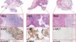
Immunohistochemical staining was performed on OSCC (n = 20), LSCC (n = 10) and PTC (n = 10), assessing CEA, c-Met, EpCAM, EGFR, integrin αvβ6 and VEGF-α. Expression was scored (0–12) using the total immunostaining score (TIS) system, and categorized into absent (TIS 0), low (TIS 1–5), moderate (TIS 6–8) or high (TIS 9–12).
Results
Integrin αvβ6 showed significant overexpression in OSCC (TIS: 12; p < 0.001) and LSCC (TIS: 8; p = 0.002), with 80% of OSCC and 90% of LSCC exhibiting moderate-high expression. Similarly, EGFR expression was moderate-high in most OSCC (87.5%; TIS: 8) and universally high in LSCC (100%; TIS: 12). In PTC, EGFR and VEGF-α expressions were low-moderate, but significantly higher than in healthy tissue (TIS: 6; p < 0.006).
Conclusion
This study highlights integrin αvβ6 and EGFR as viable FLI targets in OSCC and LSCC, especially integrin αvβ6 for tumour margin delineation. In PTC, despite lower expressions, the significant overexpression of VEGF-α, c-MET, and EGFR suggests their potential as FLI targets. Our findings support the development of tumour-targeted FLI tracers to improve surgical precision in HNC.
期刊介绍:
Molecular Diagnosis & Therapy welcomes current opinion articles on emerging or contentious issues, comprehensive narrative reviews, systematic reviews (as outlined by the PRISMA statement), original research articles (including short communications) and letters to the editor. All manuscripts are subject to peer review by international experts.
 求助内容:
求助内容: 应助结果提醒方式:


