A. Mariscal Martínez , E. Iglesias Bravo , H. Peris Alvà , P. Rodríguez Martínez , M. Luna Tomás , I. Pascual Miguel , P. Puyalto de Pablo
{"title":"用于检测乳腺癌新辅助治疗后残留病灶的对比增强乳腺 X 线照相术和磁性种子标记技术","authors":"A. Mariscal Martínez , E. Iglesias Bravo , H. Peris Alvà , P. Rodríguez Martínez , M. Luna Tomás , I. Pascual Miguel , P. Puyalto de Pablo","doi":"10.1016/j.rx.2024.04.003","DOIUrl":null,"url":null,"abstract":"<div><h3>Purpose</h3><p>Assess whether contrast-enhanced mammography (CEM) enables an evaluation of the residual size of breast tumours following neoadjuvant systemic therapy (NAST) in patients initially marked with magnetic seed.</p></div><div><h3>Materials and methods</h3><p>This single-centre prospective study was performed between March 2022 and April 2023 with patients with invasive breast carcinoma and lesional marking with magnetic seed. CEM was performed before and after NAST. The lesion size in CEM after NAST was compared to the pathological examination after surgery. Differences between sizes were evaluated and we determined the diagnostic capability indices.</p></div><div><h3>Results</h3><p>The breast lesions marked with magnetic seed were successfully localised in the preoperative stage for the 42 patients included in the study and selective surgical excision was also achieved in all cases. Tumour diameter after NAST was determined by comparing enhancement on combined CEM images from before and after NAST. The mean diameter was 13.6 mm while post-surgical pathological examination determined the mean diameter to be 12.9 mm. There were therefore no statistically significant differences between the measurements.</p></div><div><h3>Conclusions</h3><p>There is a positive correlation and similarity between CEM and pathological examination with regards to the detection of residual disease after NAST, with high specificity and positive predictive value.</p></div>","PeriodicalId":31509,"journal":{"name":"RADIOLOGIA","volume":"66 5","pages":"Pages 419-430"},"PeriodicalIF":1.1000,"publicationDate":"2024-09-01","publicationTypes":"Journal Article","fieldsOfStudy":null,"isOpenAccess":false,"openAccessPdf":"","citationCount":"0","resultStr":"{\"title\":\"Mamografía con contraste y marcaje con semilla magnética para la detección de enfermedad residual en el cáncer de mama tras tratamiento neoadyuvante\",\"authors\":\"A. Mariscal Martínez , E. Iglesias Bravo , H. Peris Alvà , P. Rodríguez Martínez , M. Luna Tomás , I. Pascual Miguel , P. Puyalto de Pablo\",\"doi\":\"10.1016/j.rx.2024.04.003\",\"DOIUrl\":null,\"url\":null,\"abstract\":\"<div><h3>Purpose</h3><p>Assess whether contrast-enhanced mammography (CEM) enables an evaluation of the residual size of breast tumours following neoadjuvant systemic therapy (NAST) in patients initially marked with magnetic seed.</p></div><div><h3>Materials and methods</h3><p>This single-centre prospective study was performed between March 2022 and April 2023 with patients with invasive breast carcinoma and lesional marking with magnetic seed. CEM was performed before and after NAST. The lesion size in CEM after NAST was compared to the pathological examination after surgery. Differences between sizes were evaluated and we determined the diagnostic capability indices.</p></div><div><h3>Results</h3><p>The breast lesions marked with magnetic seed were successfully localised in the preoperative stage for the 42 patients included in the study and selective surgical excision was also achieved in all cases. Tumour diameter after NAST was determined by comparing enhancement on combined CEM images from before and after NAST. The mean diameter was 13.6 mm while post-surgical pathological examination determined the mean diameter to be 12.9 mm. There were therefore no statistically significant differences between the measurements.</p></div><div><h3>Conclusions</h3><p>There is a positive correlation and similarity between CEM and pathological examination with regards to the detection of residual disease after NAST, with high specificity and positive predictive value.</p></div>\",\"PeriodicalId\":31509,\"journal\":{\"name\":\"RADIOLOGIA\",\"volume\":\"66 5\",\"pages\":\"Pages 419-430\"},\"PeriodicalIF\":1.1000,\"publicationDate\":\"2024-09-01\",\"publicationTypes\":\"Journal Article\",\"fieldsOfStudy\":null,\"isOpenAccess\":false,\"openAccessPdf\":\"\",\"citationCount\":\"0\",\"resultStr\":null,\"platform\":\"Semanticscholar\",\"paperid\":null,\"PeriodicalName\":\"RADIOLOGIA\",\"FirstCategoryId\":\"1085\",\"ListUrlMain\":\"https://www.sciencedirect.com/science/article/pii/S0033833824000638\",\"RegionNum\":0,\"RegionCategory\":null,\"ArticlePicture\":[],\"TitleCN\":null,\"AbstractTextCN\":null,\"PMCID\":null,\"EPubDate\":\"\",\"PubModel\":\"\",\"JCR\":\"Q3\",\"JCRName\":\"RADIOLOGY, NUCLEAR MEDICINE & MEDICAL IMAGING\",\"Score\":null,\"Total\":0}","platform":"Semanticscholar","paperid":null,"PeriodicalName":"RADIOLOGIA","FirstCategoryId":"1085","ListUrlMain":"https://www.sciencedirect.com/science/article/pii/S0033833824000638","RegionNum":0,"RegionCategory":null,"ArticlePicture":[],"TitleCN":null,"AbstractTextCN":null,"PMCID":null,"EPubDate":"","PubModel":"","JCR":"Q3","JCRName":"RADIOLOGY, NUCLEAR MEDICINE & MEDICAL IMAGING","Score":null,"Total":0}
引用次数: 0
摘要
目的 评估对比增强乳腺 X 光造影术(CEM)能否评估最初使用磁性种子标记的患者在接受新辅助系统治疗(NAST)后乳腺肿瘤的残留大小。材料与方法 这项单中心前瞻性研究于 2022 年 3 月至 2023 年 4 月间进行,研究对象为浸润性乳腺癌患者和使用磁性种子标记病灶的患者。在NAST前后均进行了CEM检查。将NAST后CEM中的病灶大小与手术后的病理检查结果进行比较。我们评估了不同大小病灶之间的差异,并确定了诊断能力指数。结果42名参与研究的患者在术前阶段均成功定位了磁性种子标记的乳腺病灶,所有病例均实现了选择性手术切除。通过比较磁种标记前后的联合 CEM 图像的增强情况,确定了磁种标记后的肿瘤直径。平均直径为 13.6 毫米,而手术后病理检查确定的平均直径为 12.9 毫米。结论CEM和病理检查在检测NAST术后残留疾病方面存在正相关性和相似性,具有较高的特异性和阳性预测值。
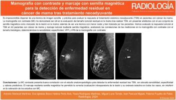
Mamografía con contraste y marcaje con semilla magnética para la detección de enfermedad residual en el cáncer de mama tras tratamiento neoadyuvante
Purpose
Assess whether contrast-enhanced mammography (CEM) enables an evaluation of the residual size of breast tumours following neoadjuvant systemic therapy (NAST) in patients initially marked with magnetic seed.
Materials and methods
This single-centre prospective study was performed between March 2022 and April 2023 with patients with invasive breast carcinoma and lesional marking with magnetic seed. CEM was performed before and after NAST. The lesion size in CEM after NAST was compared to the pathological examination after surgery. Differences between sizes were evaluated and we determined the diagnostic capability indices.
Results
The breast lesions marked with magnetic seed were successfully localised in the preoperative stage for the 42 patients included in the study and selective surgical excision was also achieved in all cases. Tumour diameter after NAST was determined by comparing enhancement on combined CEM images from before and after NAST. The mean diameter was 13.6 mm while post-surgical pathological examination determined the mean diameter to be 12.9 mm. There were therefore no statistically significant differences between the measurements.
Conclusions
There is a positive correlation and similarity between CEM and pathological examination with regards to the detection of residual disease after NAST, with high specificity and positive predictive value.
RADIOLOGIARADIOLOGY, NUCLEAR MEDICINE & MEDICAL IMAGING-
CiteScore
1.60
自引率
7.70%
发文量
105
审稿时长
52 days
期刊介绍:
La mejor revista para conocer de primera mano los originales más relevantes en la especialidad y las revisiones, casos y notas clínicas de mayor interés profesional. Además es la Publicación Oficial de la Sociedad Española de Radiología Médica.
 求助内容:
求助内容: 应助结果提醒方式:


