{"title":"阴道毛滴虫乙状结肠丝的三维细胞结构。","authors":"Sharmila Ortiz , Raphael Verdan , Marlene Benchimol","doi":"10.1016/j.jsb.2024.108127","DOIUrl":null,"url":null,"abstract":"<div><p><em>Trichomonas vaginalis</em> is a parasite protozoan that causes human trichomoniasis, a sexually transmitted infection (STI) that affects more than 156 million people worldwide. <em>T. vaginalis</em> contains an uncommon and complex cytoskeleton constituting the mastigont system, formed by several fibers and proteinaceous structures associated with basal bodies. Among these structures is the pelta-axostylar complex made of microtubules and striated filaments such as the costa and the parabasal filaments. In addition, some structures are poorly known and studied, such as the sigmoid filament and the X-filament. Here, we have isolated the <em>Trichomonas vaginalis</em> cytoskeleton and used UHR-SEM (ultra-high resolution scanning electron microscopy), tomography, immunofluorescence, immunolabeling, and backscattered electrons on SEM, negative staining to model the three-dimensional architecture and possible function of the sigmoid.</p></div>","PeriodicalId":17074,"journal":{"name":"Journal of structural biology","volume":"216 4","pages":"Article 108127"},"PeriodicalIF":2.7000,"publicationDate":"2024-09-06","publicationTypes":"Journal Article","fieldsOfStudy":null,"isOpenAccess":false,"openAccessPdf":"","citationCount":"0","resultStr":"{\"title\":\"Three-dimensional cellular architecture of the sigmoid filament in Trichomonas vaginalis\",\"authors\":\"Sharmila Ortiz , Raphael Verdan , Marlene Benchimol\",\"doi\":\"10.1016/j.jsb.2024.108127\",\"DOIUrl\":null,\"url\":null,\"abstract\":\"<div><p><em>Trichomonas vaginalis</em> is a parasite protozoan that causes human trichomoniasis, a sexually transmitted infection (STI) that affects more than 156 million people worldwide. <em>T. vaginalis</em> contains an uncommon and complex cytoskeleton constituting the mastigont system, formed by several fibers and proteinaceous structures associated with basal bodies. Among these structures is the pelta-axostylar complex made of microtubules and striated filaments such as the costa and the parabasal filaments. In addition, some structures are poorly known and studied, such as the sigmoid filament and the X-filament. Here, we have isolated the <em>Trichomonas vaginalis</em> cytoskeleton and used UHR-SEM (ultra-high resolution scanning electron microscopy), tomography, immunofluorescence, immunolabeling, and backscattered electrons on SEM, negative staining to model the three-dimensional architecture and possible function of the sigmoid.</p></div>\",\"PeriodicalId\":17074,\"journal\":{\"name\":\"Journal of structural biology\",\"volume\":\"216 4\",\"pages\":\"Article 108127\"},\"PeriodicalIF\":2.7000,\"publicationDate\":\"2024-09-06\",\"publicationTypes\":\"Journal Article\",\"fieldsOfStudy\":null,\"isOpenAccess\":false,\"openAccessPdf\":\"\",\"citationCount\":\"0\",\"resultStr\":null,\"platform\":\"Semanticscholar\",\"paperid\":null,\"PeriodicalName\":\"Journal of structural biology\",\"FirstCategoryId\":\"99\",\"ListUrlMain\":\"https://www.sciencedirect.com/science/article/pii/S1047847724000674\",\"RegionNum\":3,\"RegionCategory\":\"生物学\",\"ArticlePicture\":[],\"TitleCN\":null,\"AbstractTextCN\":null,\"PMCID\":null,\"EPubDate\":\"\",\"PubModel\":\"\",\"JCR\":\"Q3\",\"JCRName\":\"BIOCHEMISTRY & MOLECULAR BIOLOGY\",\"Score\":null,\"Total\":0}","platform":"Semanticscholar","paperid":null,"PeriodicalName":"Journal of structural biology","FirstCategoryId":"99","ListUrlMain":"https://www.sciencedirect.com/science/article/pii/S1047847724000674","RegionNum":3,"RegionCategory":"生物学","ArticlePicture":[],"TitleCN":null,"AbstractTextCN":null,"PMCID":null,"EPubDate":"","PubModel":"","JCR":"Q3","JCRName":"BIOCHEMISTRY & MOLECULAR BIOLOGY","Score":null,"Total":0}
引用次数: 0
摘要
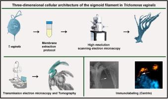
阴道毛滴虫(Trichomonas vaginalis)是一种原生寄生虫,可导致人类滴虫病,这是一种性传播感染(STI),全球有超过 1.56 亿人受到感染。阴道毛滴虫含有一种不常见的复杂细胞骨架,由与基底体相关的几种纤维和蛋白结构组成,构成马氏体系统。在这些结构中,有由微管和条状细丝(如costa和parabasal细丝)组成的pelta-axostylar复合体。此外,对一些结构的了解和研究也不多,如乙状丝和 X 丝。在这里,我们分离了阴道毛滴虫的细胞骨架,并利用超高分辨率扫描电子显微镜(UHR-SEM)、断层扫描、免疫荧光、免疫标记和扫描电子显微镜上的背散射电子、阴性染色来模拟乙状结肠的三维结构和可能的功能。
Three-dimensional cellular architecture of the sigmoid filament in Trichomonas vaginalis
Trichomonas vaginalis is a parasite protozoan that causes human trichomoniasis, a sexually transmitted infection (STI) that affects more than 156 million people worldwide. T. vaginalis contains an uncommon and complex cytoskeleton constituting the mastigont system, formed by several fibers and proteinaceous structures associated with basal bodies. Among these structures is the pelta-axostylar complex made of microtubules and striated filaments such as the costa and the parabasal filaments. In addition, some structures are poorly known and studied, such as the sigmoid filament and the X-filament. Here, we have isolated the Trichomonas vaginalis cytoskeleton and used UHR-SEM (ultra-high resolution scanning electron microscopy), tomography, immunofluorescence, immunolabeling, and backscattered electrons on SEM, negative staining to model the three-dimensional architecture and possible function of the sigmoid.
期刊介绍:
Journal of Structural Biology (JSB) has an open access mirror journal, the Journal of Structural Biology: X (JSBX), sharing the same aims and scope, editorial team, submission system and rigorous peer review. Since both journals share the same editorial system, you may submit your manuscript via either journal homepage. You will be prompted during submission (and revision) to choose in which to publish your article. The editors and reviewers are not aware of the choice you made until the article has been published online. JSB and JSBX publish papers dealing with the structural analysis of living material at every level of organization by all methods that lead to an understanding of biological function in terms of molecular and supermolecular structure.
Techniques covered include:
• Light microscopy including confocal microscopy
• All types of electron microscopy
• X-ray diffraction
• Nuclear magnetic resonance
• Scanning force microscopy, scanning probe microscopy, and tunneling microscopy
• Digital image processing
• Computational insights into structure

 求助内容:
求助内容: 应助结果提醒方式:


