{"title":"婴幼儿肾病性胱氨酸病患者的眼部受累。","authors":"Sema Üzüm, Ayşe Bozkurt Oflaz, Sadagat Guluzade, Emine Tınkır Kayıtmazbatır, Banu Bozkurt","doi":"10.4274/tjo.galenos.2024.89957","DOIUrl":null,"url":null,"abstract":"<p><p>Cystinosis is a rare autosomal recessive lysosomal storage disease associated with high mortality and morbidity rates. The most distinctive ocular manifestations of cystinosis are photophobia, tearing, and blurred vision. Herein, we assessed the ocular involvement of four patients from two families diagnosed with infantile nephropathic cystinosis using optical coherence tomography (OCT) and <i>in vivo</i> confocal microscopy (IVCM). Anterior segment OCT demonstrated multiple hyperreflective punctate deposits, and IVCM revealed needle-shaped bright crystal deposits in the corneal stroma in all patients. Three patients also had crystal deposits in the epithelium, where epithelial cell disruption was observed. Crystal deposits around the subepithelial nerve plexus were noted in some sections. In one patient, round and needle-shaped bright deposits along with inflammatory cells were observed in the limbal region of the conjunctiva. Infrared fundus images of two female siblings revealed hyperreflective crystal-like deposits around the optic disc, macula, and peripheral retina, and enhanced depth imaging OCT showed accumulation of crystals in all layers of the retina.</p>","PeriodicalId":23373,"journal":{"name":"Turkish Journal of Ophthalmology","volume":"54 4","pages":"235-239"},"PeriodicalIF":0.0000,"publicationDate":"2024-08-28","publicationTypes":"Journal Article","fieldsOfStudy":null,"isOpenAccess":false,"openAccessPdf":"https://www.ncbi.nlm.nih.gov/pmc/articles/PMC11590711/pdf/","citationCount":"0","resultStr":"{\"title\":\"Ocular Involvement in Patients with Infantile Nephropathic Cystinosis.\",\"authors\":\"Sema Üzüm, Ayşe Bozkurt Oflaz, Sadagat Guluzade, Emine Tınkır Kayıtmazbatır, Banu Bozkurt\",\"doi\":\"10.4274/tjo.galenos.2024.89957\",\"DOIUrl\":null,\"url\":null,\"abstract\":\"<p><p>Cystinosis is a rare autosomal recessive lysosomal storage disease associated with high mortality and morbidity rates. The most distinctive ocular manifestations of cystinosis are photophobia, tearing, and blurred vision. Herein, we assessed the ocular involvement of four patients from two families diagnosed with infantile nephropathic cystinosis using optical coherence tomography (OCT) and <i>in vivo</i> confocal microscopy (IVCM). Anterior segment OCT demonstrated multiple hyperreflective punctate deposits, and IVCM revealed needle-shaped bright crystal deposits in the corneal stroma in all patients. Three patients also had crystal deposits in the epithelium, where epithelial cell disruption was observed. Crystal deposits around the subepithelial nerve plexus were noted in some sections. In one patient, round and needle-shaped bright deposits along with inflammatory cells were observed in the limbal region of the conjunctiva. Infrared fundus images of two female siblings revealed hyperreflective crystal-like deposits around the optic disc, macula, and peripheral retina, and enhanced depth imaging OCT showed accumulation of crystals in all layers of the retina.</p>\",\"PeriodicalId\":23373,\"journal\":{\"name\":\"Turkish Journal of Ophthalmology\",\"volume\":\"54 4\",\"pages\":\"235-239\"},\"PeriodicalIF\":0.0000,\"publicationDate\":\"2024-08-28\",\"publicationTypes\":\"Journal Article\",\"fieldsOfStudy\":null,\"isOpenAccess\":false,\"openAccessPdf\":\"https://www.ncbi.nlm.nih.gov/pmc/articles/PMC11590711/pdf/\",\"citationCount\":\"0\",\"resultStr\":null,\"platform\":\"Semanticscholar\",\"paperid\":null,\"PeriodicalName\":\"Turkish Journal of Ophthalmology\",\"FirstCategoryId\":\"1085\",\"ListUrlMain\":\"https://doi.org/10.4274/tjo.galenos.2024.89957\",\"RegionNum\":0,\"RegionCategory\":null,\"ArticlePicture\":[],\"TitleCN\":null,\"AbstractTextCN\":null,\"PMCID\":null,\"EPubDate\":\"\",\"PubModel\":\"\",\"JCR\":\"Q3\",\"JCRName\":\"Medicine\",\"Score\":null,\"Total\":0}","platform":"Semanticscholar","paperid":null,"PeriodicalName":"Turkish Journal of Ophthalmology","FirstCategoryId":"1085","ListUrlMain":"https://doi.org/10.4274/tjo.galenos.2024.89957","RegionNum":0,"RegionCategory":null,"ArticlePicture":[],"TitleCN":null,"AbstractTextCN":null,"PMCID":null,"EPubDate":"","PubModel":"","JCR":"Q3","JCRName":"Medicine","Score":null,"Total":0}
引用次数: 0
摘要
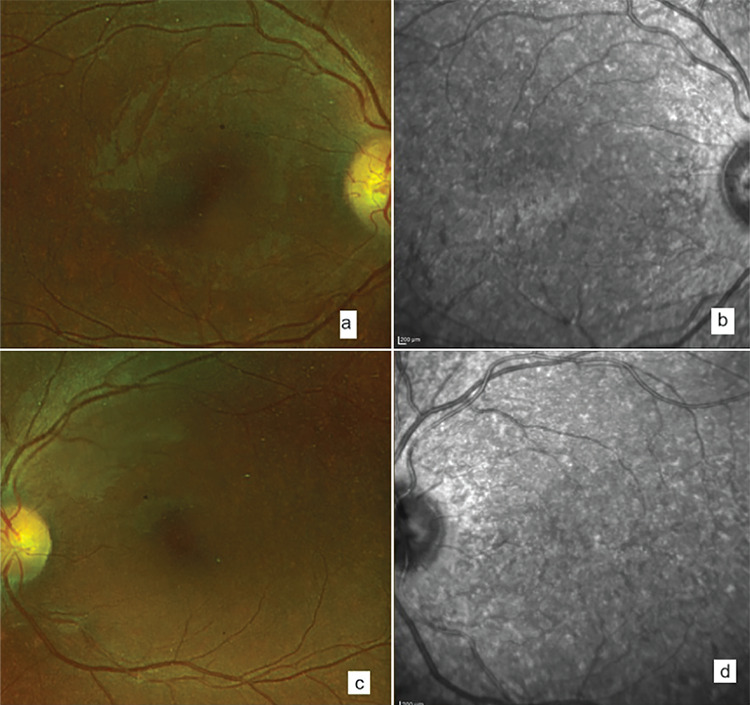
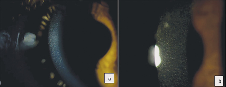
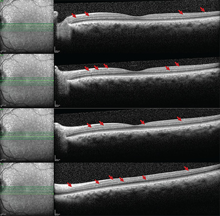
胱氨酸病是一种罕见的常染色体隐性溶酶体储积病,死亡率和发病率都很高。胱氨酸病最明显的眼部表现是畏光、流泪和视力模糊。在此,我们使用光学相干断层扫描(OCT)和活体共聚焦显微镜(IVCM)评估了来自两个家族的四名被诊断为婴幼儿肾病性胱氨酸病的患者的眼部受累情况。前段光学相干断层扫描(OCT)显示出多个高反射点状沉积物,IVCM 显示所有患者的角膜基质中都有针状明亮晶体沉积物。三名患者的上皮细胞也有晶体沉积,并观察到上皮细胞破坏。在一些切片中发现上皮下神经丛周围有晶体沉积。一名患者的结膜边缘区域观察到圆形和针状的明亮沉积物以及炎性细胞。两名女性兄弟姐妹的红外眼底图像显示,视盘、黄斑和周边视网膜周围有高反射晶体样沉积物,增强深度成像 OCT 显示视网膜各层均有晶体积聚。
Ocular Involvement in Patients with Infantile Nephropathic Cystinosis.
Cystinosis is a rare autosomal recessive lysosomal storage disease associated with high mortality and morbidity rates. The most distinctive ocular manifestations of cystinosis are photophobia, tearing, and blurred vision. Herein, we assessed the ocular involvement of four patients from two families diagnosed with infantile nephropathic cystinosis using optical coherence tomography (OCT) and in vivo confocal microscopy (IVCM). Anterior segment OCT demonstrated multiple hyperreflective punctate deposits, and IVCM revealed needle-shaped bright crystal deposits in the corneal stroma in all patients. Three patients also had crystal deposits in the epithelium, where epithelial cell disruption was observed. Crystal deposits around the subepithelial nerve plexus were noted in some sections. In one patient, round and needle-shaped bright deposits along with inflammatory cells were observed in the limbal region of the conjunctiva. Infrared fundus images of two female siblings revealed hyperreflective crystal-like deposits around the optic disc, macula, and peripheral retina, and enhanced depth imaging OCT showed accumulation of crystals in all layers of the retina.
期刊介绍:
The Turkish Journal of Ophthalmology (TJO) is the only scientific periodical publication of the Turkish Ophthalmological Association and has been published since January 1929. In its early years, the journal was published in Turkish and French. Although there were temporary interruptions in the publication of the journal due to various challenges, the Turkish Journal of Ophthalmology has been published continually from 1971 to the present. The target audience includes specialists and physicians in training in ophthalmology in all relevant disciplines.

 求助内容:
求助内容: 应助结果提醒方式:


