Yeyang Xian, Jie Liu, Mengxuan Dai, Wensheng Zhang, Minye He, Zhengnong Wei, Yutao Jiang, Shiyong Le, Zhuoang Lin, Shuai Tang, Yunfei Zhou, Liming Dong, Jinzheng Liang, Jie Zhang, Liang Wang
{"title":"急性脊髓损伤后,小胶质细胞通过 VEGF-C/VEGFR3 依赖性自噬和极化促进脊髓周围淋巴管生成","authors":"Yeyang Xian, Jie Liu, Mengxuan Dai, Wensheng Zhang, Minye He, Zhengnong Wei, Yutao Jiang, Shiyong Le, Zhuoang Lin, Shuai Tang, Yunfei Zhou, Liming Dong, Jinzheng Liang, Jie Zhang, Liang Wang","doi":"10.1007/s12035-024-04437-5","DOIUrl":null,"url":null,"abstract":"<p><p>Reducing secondary injury is a key focus in the field of spinal cord injury (SCI). Recent studies have revealed the role of lymphangiogenesis in reducing secondary damage to central nerve. However, the mechanism of lymphangiogenesis is not yet clear. Macrophages have been shown to play an important role in peripheral tissue lymphangiogenesis. Microglia is believed to play a role similar to macrophages in the central nervous system (CNS); we hypothesized that there was a close relationship between microglia and central nerve system lymphangiogenesis. Herein, we used an in vivo model of SCI to explored the relationship between microglia and spinal cord lymphangiogenesis and further investigated the polarization of microglia and its role in promoting spinal cord lymphangiogenesis by a series of in vitro experiments. The current study elucidated for the first time the relationship between microglia and lymphangiogenesis around the spinal cord after SCI. Classical activated (M1) microglia can promote lymphangiogenesis by secreting VEGF-C which further increases polarization and secretion of lymphatic growth factor by activating VEGFR3. The VEGF-C/VEGFR3 pathway activation downregulates microglia autophagy, thereby regulating the microglia phenotype. These results indicate that M1 microglia promote lymphangiogenesis after SCI, and activated VEGF-C/VEGFR3 signaling promotes M1 microglia polarization by inhibiting autophagy, thereby facilitates lymphangiogenesis.</p>","PeriodicalId":18762,"journal":{"name":"Molecular Neurobiology","volume":" ","pages":"2740-2755"},"PeriodicalIF":4.6000,"publicationDate":"2025-03-01","publicationTypes":"Journal Article","fieldsOfStudy":null,"isOpenAccess":false,"openAccessPdf":"","citationCount":"0","resultStr":"{\"title\":\"Microglia Promote Lymphangiogenesis Around the Spinal Cord Through VEGF-C/VEGFR3-Dependent Autophagy and Polarization After Acute Spinal Cord Injury.\",\"authors\":\"Yeyang Xian, Jie Liu, Mengxuan Dai, Wensheng Zhang, Minye He, Zhengnong Wei, Yutao Jiang, Shiyong Le, Zhuoang Lin, Shuai Tang, Yunfei Zhou, Liming Dong, Jinzheng Liang, Jie Zhang, Liang Wang\",\"doi\":\"10.1007/s12035-024-04437-5\",\"DOIUrl\":null,\"url\":null,\"abstract\":\"<p><p>Reducing secondary injury is a key focus in the field of spinal cord injury (SCI). Recent studies have revealed the role of lymphangiogenesis in reducing secondary damage to central nerve. However, the mechanism of lymphangiogenesis is not yet clear. Macrophages have been shown to play an important role in peripheral tissue lymphangiogenesis. Microglia is believed to play a role similar to macrophages in the central nervous system (CNS); we hypothesized that there was a close relationship between microglia and central nerve system lymphangiogenesis. Herein, we used an in vivo model of SCI to explored the relationship between microglia and spinal cord lymphangiogenesis and further investigated the polarization of microglia and its role in promoting spinal cord lymphangiogenesis by a series of in vitro experiments. The current study elucidated for the first time the relationship between microglia and lymphangiogenesis around the spinal cord after SCI. Classical activated (M1) microglia can promote lymphangiogenesis by secreting VEGF-C which further increases polarization and secretion of lymphatic growth factor by activating VEGFR3. The VEGF-C/VEGFR3 pathway activation downregulates microglia autophagy, thereby regulating the microglia phenotype. These results indicate that M1 microglia promote lymphangiogenesis after SCI, and activated VEGF-C/VEGFR3 signaling promotes M1 microglia polarization by inhibiting autophagy, thereby facilitates lymphangiogenesis.</p>\",\"PeriodicalId\":18762,\"journal\":{\"name\":\"Molecular Neurobiology\",\"volume\":\" \",\"pages\":\"2740-2755\"},\"PeriodicalIF\":4.6000,\"publicationDate\":\"2025-03-01\",\"publicationTypes\":\"Journal Article\",\"fieldsOfStudy\":null,\"isOpenAccess\":false,\"openAccessPdf\":\"\",\"citationCount\":\"0\",\"resultStr\":null,\"platform\":\"Semanticscholar\",\"paperid\":null,\"PeriodicalName\":\"Molecular Neurobiology\",\"FirstCategoryId\":\"3\",\"ListUrlMain\":\"https://doi.org/10.1007/s12035-024-04437-5\",\"RegionNum\":2,\"RegionCategory\":\"医学\",\"ArticlePicture\":[],\"TitleCN\":null,\"AbstractTextCN\":null,\"PMCID\":null,\"EPubDate\":\"2024/8/19 0:00:00\",\"PubModel\":\"Epub\",\"JCR\":\"Q1\",\"JCRName\":\"NEUROSCIENCES\",\"Score\":null,\"Total\":0}","platform":"Semanticscholar","paperid":null,"PeriodicalName":"Molecular Neurobiology","FirstCategoryId":"3","ListUrlMain":"https://doi.org/10.1007/s12035-024-04437-5","RegionNum":2,"RegionCategory":"医学","ArticlePicture":[],"TitleCN":null,"AbstractTextCN":null,"PMCID":null,"EPubDate":"2024/8/19 0:00:00","PubModel":"Epub","JCR":"Q1","JCRName":"NEUROSCIENCES","Score":null,"Total":0}
Microglia Promote Lymphangiogenesis Around the Spinal Cord Through VEGF-C/VEGFR3-Dependent Autophagy and Polarization After Acute Spinal Cord Injury.
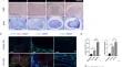
Reducing secondary injury is a key focus in the field of spinal cord injury (SCI). Recent studies have revealed the role of lymphangiogenesis in reducing secondary damage to central nerve. However, the mechanism of lymphangiogenesis is not yet clear. Macrophages have been shown to play an important role in peripheral tissue lymphangiogenesis. Microglia is believed to play a role similar to macrophages in the central nervous system (CNS); we hypothesized that there was a close relationship between microglia and central nerve system lymphangiogenesis. Herein, we used an in vivo model of SCI to explored the relationship between microglia and spinal cord lymphangiogenesis and further investigated the polarization of microglia and its role in promoting spinal cord lymphangiogenesis by a series of in vitro experiments. The current study elucidated for the first time the relationship between microglia and lymphangiogenesis around the spinal cord after SCI. Classical activated (M1) microglia can promote lymphangiogenesis by secreting VEGF-C which further increases polarization and secretion of lymphatic growth factor by activating VEGFR3. The VEGF-C/VEGFR3 pathway activation downregulates microglia autophagy, thereby regulating the microglia phenotype. These results indicate that M1 microglia promote lymphangiogenesis after SCI, and activated VEGF-C/VEGFR3 signaling promotes M1 microglia polarization by inhibiting autophagy, thereby facilitates lymphangiogenesis.
期刊介绍:
Molecular Neurobiology is an exciting journal for neuroscientists needing to stay in close touch with progress at the forefront of molecular brain research today. It is an especially important periodical for graduate students and "postdocs," specifically designed to synthesize and critically assess research trends for all neuroscientists hoping to stay active at the cutting edge of this dramatically developing area. This journal has proven to be crucial in departmental libraries, serving as essential reading for every committed neuroscientist who is striving to keep abreast of all rapid developments in a forefront field. Most recent significant advances in experimental and clinical neuroscience have been occurring at the molecular level. Until now, there has been no journal devoted to looking closely at this fragmented literature in a critical, coherent fashion. Each submission is thoroughly analyzed by scientists and clinicians internationally renowned for their special competence in the areas treated.
 求助内容:
求助内容: 应助结果提醒方式:


