{"title":"用于胃癌检测的组织病理学图像分析:深度学习和 catboost 混合方法","authors":"Danial Khayatian, Alireza Maleki, Hamid Nasiri, Morteza Dorrigiv","doi":"10.1007/s11042-024-19816-2","DOIUrl":null,"url":null,"abstract":"<p>Since gastric cancer is growing fast, accurate and prompt diagnosis is essential, utilizing computer-aided diagnosis (CAD) systems is an efficient way to achieve this goal. Using methods related to computer vision enables more accurate predictions and faster diagnosis, leading to timely treatment. CAD systems can categorize photos effectively using deep learning techniques based on image analysis and classification. Accurate and timely classification of histopathology images is critical for enabling immediate treatment strategies, but remains challenging. We propose a hybrid deep learning and gradient-boosting approach that achieves high accuracy in classifying gastric histopathology images. This approach examines two classifiers for six networks known as pre-trained models to extract features. Extracted features will be fed to the classifiers separately. The inputs are gastric histopathological images. The GasHisSDB dataset provides these inputs containing histopathology gastric images in three 80px, 120px, and 160px cropping sizes. According to these achievements and experiments, we proposed the final method, which combines the EfficientNetV2B0 model to extract features from the images and then classify them using the CatBoost classifier. The results based on the accuracy score are 89.7%, 93.1%, and 93.9% in 80px, 120px, and 160px cropping sizes, respectively. Additional metrics including precision, recall, and F1-scores were above 0.9, demonstrating strong performance across various evaluation criteria. In another way, to approve and see the model efficiency, the GradCAM algorithm was implemented. Visualization via Grad-CAM illustrated discriminative regions identified by the model, confirming focused learning on histologically relevant features. The consistent accuracy and reliable detections across diverse evaluation metrics substantiate the robustness of the proposed deep learning and gradient-boosting approach for gastric cancer screening from histopathology images. For this purpose, two types of outputs (The heat map and the GradCAM output) are provided. Additionally, t-SNE visualization showed a clear clustering of normal and abnormal cases after EfficientNetV2B0 feature extraction. The cross-validation and visualizations provide further evidence of generalizability and focused learning of meaningful pathology features for gastric cancer screening from histopathology images.</p>","PeriodicalId":18770,"journal":{"name":"Multimedia Tools and Applications","volume":"24 1","pages":""},"PeriodicalIF":3.0000,"publicationDate":"2024-08-07","publicationTypes":"Journal Article","fieldsOfStudy":null,"isOpenAccess":false,"openAccessPdf":"","citationCount":"0","resultStr":"{\"title\":\"Histopathology image analysis for gastric cancer detection: a hybrid deep learning and catboost approach\",\"authors\":\"Danial Khayatian, Alireza Maleki, Hamid Nasiri, Morteza Dorrigiv\",\"doi\":\"10.1007/s11042-024-19816-2\",\"DOIUrl\":null,\"url\":null,\"abstract\":\"<p>Since gastric cancer is growing fast, accurate and prompt diagnosis is essential, utilizing computer-aided diagnosis (CAD) systems is an efficient way to achieve this goal. Using methods related to computer vision enables more accurate predictions and faster diagnosis, leading to timely treatment. CAD systems can categorize photos effectively using deep learning techniques based on image analysis and classification. Accurate and timely classification of histopathology images is critical for enabling immediate treatment strategies, but remains challenging. We propose a hybrid deep learning and gradient-boosting approach that achieves high accuracy in classifying gastric histopathology images. This approach examines two classifiers for six networks known as pre-trained models to extract features. Extracted features will be fed to the classifiers separately. The inputs are gastric histopathological images. The GasHisSDB dataset provides these inputs containing histopathology gastric images in three 80px, 120px, and 160px cropping sizes. According to these achievements and experiments, we proposed the final method, which combines the EfficientNetV2B0 model to extract features from the images and then classify them using the CatBoost classifier. The results based on the accuracy score are 89.7%, 93.1%, and 93.9% in 80px, 120px, and 160px cropping sizes, respectively. Additional metrics including precision, recall, and F1-scores were above 0.9, demonstrating strong performance across various evaluation criteria. In another way, to approve and see the model efficiency, the GradCAM algorithm was implemented. Visualization via Grad-CAM illustrated discriminative regions identified by the model, confirming focused learning on histologically relevant features. The consistent accuracy and reliable detections across diverse evaluation metrics substantiate the robustness of the proposed deep learning and gradient-boosting approach for gastric cancer screening from histopathology images. For this purpose, two types of outputs (The heat map and the GradCAM output) are provided. Additionally, t-SNE visualization showed a clear clustering of normal and abnormal cases after EfficientNetV2B0 feature extraction. The cross-validation and visualizations provide further evidence of generalizability and focused learning of meaningful pathology features for gastric cancer screening from histopathology images.</p>\",\"PeriodicalId\":18770,\"journal\":{\"name\":\"Multimedia Tools and Applications\",\"volume\":\"24 1\",\"pages\":\"\"},\"PeriodicalIF\":3.0000,\"publicationDate\":\"2024-08-07\",\"publicationTypes\":\"Journal Article\",\"fieldsOfStudy\":null,\"isOpenAccess\":false,\"openAccessPdf\":\"\",\"citationCount\":\"0\",\"resultStr\":null,\"platform\":\"Semanticscholar\",\"paperid\":null,\"PeriodicalName\":\"Multimedia Tools and Applications\",\"FirstCategoryId\":\"94\",\"ListUrlMain\":\"https://doi.org/10.1007/s11042-024-19816-2\",\"RegionNum\":4,\"RegionCategory\":\"计算机科学\",\"ArticlePicture\":[],\"TitleCN\":null,\"AbstractTextCN\":null,\"PMCID\":null,\"EPubDate\":\"\",\"PubModel\":\"\",\"JCR\":\"Q2\",\"JCRName\":\"COMPUTER SCIENCE, INFORMATION SYSTEMS\",\"Score\":null,\"Total\":0}","platform":"Semanticscholar","paperid":null,"PeriodicalName":"Multimedia Tools and Applications","FirstCategoryId":"94","ListUrlMain":"https://doi.org/10.1007/s11042-024-19816-2","RegionNum":4,"RegionCategory":"计算机科学","ArticlePicture":[],"TitleCN":null,"AbstractTextCN":null,"PMCID":null,"EPubDate":"","PubModel":"","JCR":"Q2","JCRName":"COMPUTER SCIENCE, INFORMATION SYSTEMS","Score":null,"Total":0}
Histopathology image analysis for gastric cancer detection: a hybrid deep learning and catboost approach
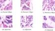
Since gastric cancer is growing fast, accurate and prompt diagnosis is essential, utilizing computer-aided diagnosis (CAD) systems is an efficient way to achieve this goal. Using methods related to computer vision enables more accurate predictions and faster diagnosis, leading to timely treatment. CAD systems can categorize photos effectively using deep learning techniques based on image analysis and classification. Accurate and timely classification of histopathology images is critical for enabling immediate treatment strategies, but remains challenging. We propose a hybrid deep learning and gradient-boosting approach that achieves high accuracy in classifying gastric histopathology images. This approach examines two classifiers for six networks known as pre-trained models to extract features. Extracted features will be fed to the classifiers separately. The inputs are gastric histopathological images. The GasHisSDB dataset provides these inputs containing histopathology gastric images in three 80px, 120px, and 160px cropping sizes. According to these achievements and experiments, we proposed the final method, which combines the EfficientNetV2B0 model to extract features from the images and then classify them using the CatBoost classifier. The results based on the accuracy score are 89.7%, 93.1%, and 93.9% in 80px, 120px, and 160px cropping sizes, respectively. Additional metrics including precision, recall, and F1-scores were above 0.9, demonstrating strong performance across various evaluation criteria. In another way, to approve and see the model efficiency, the GradCAM algorithm was implemented. Visualization via Grad-CAM illustrated discriminative regions identified by the model, confirming focused learning on histologically relevant features. The consistent accuracy and reliable detections across diverse evaluation metrics substantiate the robustness of the proposed deep learning and gradient-boosting approach for gastric cancer screening from histopathology images. For this purpose, two types of outputs (The heat map and the GradCAM output) are provided. Additionally, t-SNE visualization showed a clear clustering of normal and abnormal cases after EfficientNetV2B0 feature extraction. The cross-validation and visualizations provide further evidence of generalizability and focused learning of meaningful pathology features for gastric cancer screening from histopathology images.
期刊介绍:
Multimedia Tools and Applications publishes original research articles on multimedia development and system support tools as well as case studies of multimedia applications. It also features experimental and survey articles. The journal is intended for academics, practitioners, scientists and engineers who are involved in multimedia system research, design and applications. All papers are peer reviewed.
Specific areas of interest include:
- Multimedia Tools:
- Multimedia Applications:
- Prototype multimedia systems and platforms
 求助内容:
求助内容: 应助结果提醒方式:


