Antonio Ieni, Cristina Pizzimenti, Vincenzo Fiorentino, Mariausilia Franchina, Antonino Germanò, Giovanni Raffa, Maurizio Martini, Guido Fadda, Giovanni Tuccari
{"title":"人低度和高度颅内脑膜瘤中 p62/SQSTM1/Sequestosome-1 的免疫组化特征","authors":"Antonio Ieni, Cristina Pizzimenti, Vincenzo Fiorentino, Mariausilia Franchina, Antonino Germanò, Giovanni Raffa, Maurizio Martini, Guido Fadda, Giovanni Tuccari","doi":"10.1155/2024/5573892","DOIUrl":null,"url":null,"abstract":"<p><p>Among autophagic-related proteins, p62/SQSTM1/Sequestosome-1 represents a relevant actor in cellular proliferation and neoplastic growth. Although, recently, p62 expression has been analyzed in different neurodegenerative and glial neoplastic diseases, no available information have been reported in meningiomas, which have an high epidemiological relevance being the second most common category of intracranial tumors after gliomas. Generally meningiomas have a benign behavior, but their recurrence is not uncommon mainly when atypical or anaplastic varieties occur. However, intranuclear vacuoles have been ultrastructurally observed in meningiomas, and they were labelled by p62 antibodies. Therefore, in the present study, we have investigated p62 immunohistochemical pattern in a cohort of 133 cases representative of low- and high-grade meningiomas, to verify if p62 expression may be related to clinicopathological data, thus achieving a potential prognostic role. The p62 immunoexpression was frequently found in the nucleus and cytoplasm of neoplastic elements, and utilizing an intensity-distribution score, 55 (41.3%) cases were considered as high expressors while 78 (58.7%) cases were instead recorded as low expressors. Fifteen cases exhibited recurrences of the disease, 14 of which were codified as high expressors. Moreover, a direct relationship between p62 and Mib-1 immunoexpression as well as between p62 and neoplastic grade have been documented. Finally, we suggest that impaired autophagic flux with an increase in p62 expression may be involved in the activation of NRF2 also contributing in the development of recurrence in meningioma patients.</p>","PeriodicalId":49326,"journal":{"name":"Analytical Cellular Pathology","volume":"2024 ","pages":"5573892"},"PeriodicalIF":2.7000,"publicationDate":"2024-08-02","publicationTypes":"Journal Article","fieldsOfStudy":null,"isOpenAccess":false,"openAccessPdf":"https://www.ncbi.nlm.nih.gov/pmc/articles/PMC11315968/pdf/","citationCount":"0","resultStr":"{\"title\":\"Immunohistochemical Profile of p62/SQSTM1/Sequestosome-1 in Human Low- and High-Grade Intracranial Meningiomas.\",\"authors\":\"Antonio Ieni, Cristina Pizzimenti, Vincenzo Fiorentino, Mariausilia Franchina, Antonino Germanò, Giovanni Raffa, Maurizio Martini, Guido Fadda, Giovanni Tuccari\",\"doi\":\"10.1155/2024/5573892\",\"DOIUrl\":null,\"url\":null,\"abstract\":\"<p><p>Among autophagic-related proteins, p62/SQSTM1/Sequestosome-1 represents a relevant actor in cellular proliferation and neoplastic growth. Although, recently, p62 expression has been analyzed in different neurodegenerative and glial neoplastic diseases, no available information have been reported in meningiomas, which have an high epidemiological relevance being the second most common category of intracranial tumors after gliomas. Generally meningiomas have a benign behavior, but their recurrence is not uncommon mainly when atypical or anaplastic varieties occur. However, intranuclear vacuoles have been ultrastructurally observed in meningiomas, and they were labelled by p62 antibodies. Therefore, in the present study, we have investigated p62 immunohistochemical pattern in a cohort of 133 cases representative of low- and high-grade meningiomas, to verify if p62 expression may be related to clinicopathological data, thus achieving a potential prognostic role. The p62 immunoexpression was frequently found in the nucleus and cytoplasm of neoplastic elements, and utilizing an intensity-distribution score, 55 (41.3%) cases were considered as high expressors while 78 (58.7%) cases were instead recorded as low expressors. Fifteen cases exhibited recurrences of the disease, 14 of which were codified as high expressors. Moreover, a direct relationship between p62 and Mib-1 immunoexpression as well as between p62 and neoplastic grade have been documented. Finally, we suggest that impaired autophagic flux with an increase in p62 expression may be involved in the activation of NRF2 also contributing in the development of recurrence in meningioma patients.</p>\",\"PeriodicalId\":49326,\"journal\":{\"name\":\"Analytical Cellular Pathology\",\"volume\":\"2024 \",\"pages\":\"5573892\"},\"PeriodicalIF\":2.7000,\"publicationDate\":\"2024-08-02\",\"publicationTypes\":\"Journal Article\",\"fieldsOfStudy\":null,\"isOpenAccess\":false,\"openAccessPdf\":\"https://www.ncbi.nlm.nih.gov/pmc/articles/PMC11315968/pdf/\",\"citationCount\":\"0\",\"resultStr\":null,\"platform\":\"Semanticscholar\",\"paperid\":null,\"PeriodicalName\":\"Analytical Cellular Pathology\",\"FirstCategoryId\":\"3\",\"ListUrlMain\":\"https://doi.org/10.1155/2024/5573892\",\"RegionNum\":4,\"RegionCategory\":\"医学\",\"ArticlePicture\":[],\"TitleCN\":null,\"AbstractTextCN\":null,\"PMCID\":null,\"EPubDate\":\"2024/1/1 0:00:00\",\"PubModel\":\"eCollection\",\"JCR\":\"Q3\",\"JCRName\":\"CELL BIOLOGY\",\"Score\":null,\"Total\":0}","platform":"Semanticscholar","paperid":null,"PeriodicalName":"Analytical Cellular Pathology","FirstCategoryId":"3","ListUrlMain":"https://doi.org/10.1155/2024/5573892","RegionNum":4,"RegionCategory":"医学","ArticlePicture":[],"TitleCN":null,"AbstractTextCN":null,"PMCID":null,"EPubDate":"2024/1/1 0:00:00","PubModel":"eCollection","JCR":"Q3","JCRName":"CELL BIOLOGY","Score":null,"Total":0}
Immunohistochemical Profile of p62/SQSTM1/Sequestosome-1 in Human Low- and High-Grade Intracranial Meningiomas.
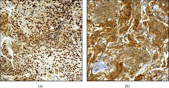
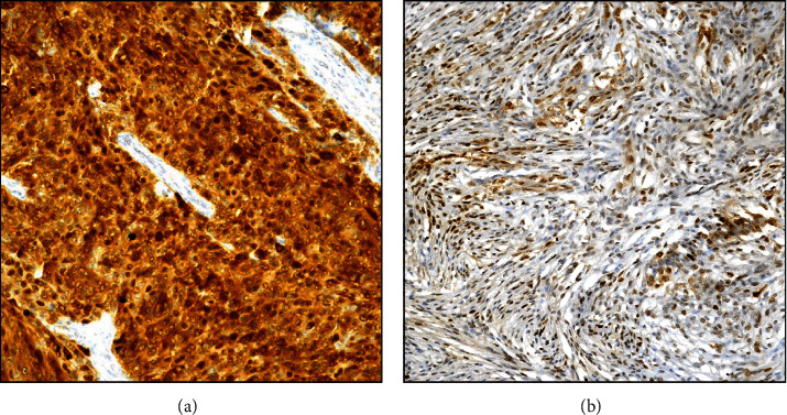
Among autophagic-related proteins, p62/SQSTM1/Sequestosome-1 represents a relevant actor in cellular proliferation and neoplastic growth. Although, recently, p62 expression has been analyzed in different neurodegenerative and glial neoplastic diseases, no available information have been reported in meningiomas, which have an high epidemiological relevance being the second most common category of intracranial tumors after gliomas. Generally meningiomas have a benign behavior, but their recurrence is not uncommon mainly when atypical or anaplastic varieties occur. However, intranuclear vacuoles have been ultrastructurally observed in meningiomas, and they were labelled by p62 antibodies. Therefore, in the present study, we have investigated p62 immunohistochemical pattern in a cohort of 133 cases representative of low- and high-grade meningiomas, to verify if p62 expression may be related to clinicopathological data, thus achieving a potential prognostic role. The p62 immunoexpression was frequently found in the nucleus and cytoplasm of neoplastic elements, and utilizing an intensity-distribution score, 55 (41.3%) cases were considered as high expressors while 78 (58.7%) cases were instead recorded as low expressors. Fifteen cases exhibited recurrences of the disease, 14 of which were codified as high expressors. Moreover, a direct relationship between p62 and Mib-1 immunoexpression as well as between p62 and neoplastic grade have been documented. Finally, we suggest that impaired autophagic flux with an increase in p62 expression may be involved in the activation of NRF2 also contributing in the development of recurrence in meningioma patients.
期刊介绍:
Analytical Cellular Pathology is a peer-reviewed, Open Access journal that provides a forum for scientists, medical practitioners and pathologists working in the area of cellular pathology. The journal publishes original research articles, review articles, and clinical studies related to cytology, carcinogenesis, cell receptors, biomarkers, diagnostic pathology, immunopathology, and hematology.
 求助内容:
求助内容: 应助结果提醒方式:


