{"title":"牙线夹牵引辅助内镜超声引导肝胃造口术,用于肝内胆管结石碎裂和清除。","authors":"Jia-Yi Ma, Zhen-Dong Jin, Kai-Xuan Wang","doi":"10.1111/den.14901","DOIUrl":null,"url":null,"abstract":"<p>A 62-year-old man who had undergone Roux-en-Y hepaticojejunostomy because of choledochal cyst presented with jaundice. Magnetic resonance cholangiopancreatography revealed obstructive stones in left intrahepatic duct (Fig. 1).</p><p>Double-ballon enteroscope-assisted endoscopic retrograde cholangiopancreatography was initially attempted but failed to reach anastomosis. Endoscopic ultrasound-guided hepaticogastrostomy (HGS) was alternatively performed (Fig. 2a). Cholangiography confirmed multiple intrahepatic bile duct stones and a 2 cm length anastomotic stricture. After dilatation of the stomach wall and anastomotic stricture, a 10 × 80 mm fully covered self-expanding metal stent (FCSEMS) was deployed.</p><p>One week later when the FCSEMS was assumed to be fully expanded (Fig. 2b), transluminal stone removal was attempted. With the concern of stent migration during stone removal, a clip and dental floss was used for FCSEMS traction and fixation (Fig. 2c). SpyGlass-guided laser lithotripsy and stone extraction was performed through the endoscopic ultrasound-guided HGS route (Fig. 2d). A coaxial double-pigtail plastic stent was inserted through the FCSEMS to function as an anchor. No postoperative adverse event was observed. Jaundice was rapidly relieved (Video S1).</p><p>Removal of left intrahepatic duct stones via the HGS route has been described in sporadic cases.<span><sup>1</sup></span> Three highlights of this case are specifically introduced as follows. First, given that the stone's size exceeded the width of intrahepatic bile duct and the diameter of metal stent when it was not fully expanded, adequate stone removal cannot be accomplished in a single session. An HGS route must first be established. Lithotripsy was then carried out. Second, to decrease the risk of stent migration during accessory devices pass-by, we borrowed the method of dental floss traction applied in endoscopic submucosal dissection.<span><sup>2</sup></span> Last, anastomotic stenosis is difficult to alleviate from a single dilation. With the help of the HGS route, double-ballon enteroscope-assisted endoscopic retrograde cholangiopancreatography can be easily performed by rendezvous technique in the subsequent session and the transgastric stent can be removed thereby.</p><p>Authors declare no conflict of interest for this article.</p><p>This study was supported by the National Natural Science Foundation of China (Grant No. 82070663 [J.Y.M.]) and Specific Research Fund of The Innovation Platform for Academicians of Hainan Province (Grant No. YSPTZX202029 [K.X.W.]).</p>","PeriodicalId":159,"journal":{"name":"Digestive Endoscopy","volume":"36 11","pages":"1284-1285"},"PeriodicalIF":5.0000,"publicationDate":"2024-08-09","publicationTypes":"Journal Article","fieldsOfStudy":null,"isOpenAccess":false,"openAccessPdf":"https://onlinelibrary.wiley.com/doi/epdf/10.1111/den.14901","citationCount":"0","resultStr":"{\"title\":\"Dental floss clip traction-assisted endoscopic ultrasound-guided hepaticogastrostomy for transluminal intrahepatic bile duct stone fragmentation and removal\",\"authors\":\"Jia-Yi Ma, Zhen-Dong Jin, Kai-Xuan Wang\",\"doi\":\"10.1111/den.14901\",\"DOIUrl\":null,\"url\":null,\"abstract\":\"<p>A 62-year-old man who had undergone Roux-en-Y hepaticojejunostomy because of choledochal cyst presented with jaundice. Magnetic resonance cholangiopancreatography revealed obstructive stones in left intrahepatic duct (Fig. 1).</p><p>Double-ballon enteroscope-assisted endoscopic retrograde cholangiopancreatography was initially attempted but failed to reach anastomosis. Endoscopic ultrasound-guided hepaticogastrostomy (HGS) was alternatively performed (Fig. 2a). Cholangiography confirmed multiple intrahepatic bile duct stones and a 2 cm length anastomotic stricture. After dilatation of the stomach wall and anastomotic stricture, a 10 × 80 mm fully covered self-expanding metal stent (FCSEMS) was deployed.</p><p>One week later when the FCSEMS was assumed to be fully expanded (Fig. 2b), transluminal stone removal was attempted. With the concern of stent migration during stone removal, a clip and dental floss was used for FCSEMS traction and fixation (Fig. 2c). SpyGlass-guided laser lithotripsy and stone extraction was performed through the endoscopic ultrasound-guided HGS route (Fig. 2d). A coaxial double-pigtail plastic stent was inserted through the FCSEMS to function as an anchor. No postoperative adverse event was observed. Jaundice was rapidly relieved (Video S1).</p><p>Removal of left intrahepatic duct stones via the HGS route has been described in sporadic cases.<span><sup>1</sup></span> Three highlights of this case are specifically introduced as follows. First, given that the stone's size exceeded the width of intrahepatic bile duct and the diameter of metal stent when it was not fully expanded, adequate stone removal cannot be accomplished in a single session. An HGS route must first be established. Lithotripsy was then carried out. Second, to decrease the risk of stent migration during accessory devices pass-by, we borrowed the method of dental floss traction applied in endoscopic submucosal dissection.<span><sup>2</sup></span> Last, anastomotic stenosis is difficult to alleviate from a single dilation. With the help of the HGS route, double-ballon enteroscope-assisted endoscopic retrograde cholangiopancreatography can be easily performed by rendezvous technique in the subsequent session and the transgastric stent can be removed thereby.</p><p>Authors declare no conflict of interest for this article.</p><p>This study was supported by the National Natural Science Foundation of China (Grant No. 82070663 [J.Y.M.]) and Specific Research Fund of The Innovation Platform for Academicians of Hainan Province (Grant No. YSPTZX202029 [K.X.W.]).</p>\",\"PeriodicalId\":159,\"journal\":{\"name\":\"Digestive Endoscopy\",\"volume\":\"36 11\",\"pages\":\"1284-1285\"},\"PeriodicalIF\":5.0000,\"publicationDate\":\"2024-08-09\",\"publicationTypes\":\"Journal Article\",\"fieldsOfStudy\":null,\"isOpenAccess\":false,\"openAccessPdf\":\"https://onlinelibrary.wiley.com/doi/epdf/10.1111/den.14901\",\"citationCount\":\"0\",\"resultStr\":null,\"platform\":\"Semanticscholar\",\"paperid\":null,\"PeriodicalName\":\"Digestive Endoscopy\",\"FirstCategoryId\":\"3\",\"ListUrlMain\":\"https://onlinelibrary.wiley.com/doi/10.1111/den.14901\",\"RegionNum\":2,\"RegionCategory\":\"医学\",\"ArticlePicture\":[],\"TitleCN\":null,\"AbstractTextCN\":null,\"PMCID\":null,\"EPubDate\":\"\",\"PubModel\":\"\",\"JCR\":\"Q1\",\"JCRName\":\"GASTROENTEROLOGY & HEPATOLOGY\",\"Score\":null,\"Total\":0}","platform":"Semanticscholar","paperid":null,"PeriodicalName":"Digestive Endoscopy","FirstCategoryId":"3","ListUrlMain":"https://onlinelibrary.wiley.com/doi/10.1111/den.14901","RegionNum":2,"RegionCategory":"医学","ArticlePicture":[],"TitleCN":null,"AbstractTextCN":null,"PMCID":null,"EPubDate":"","PubModel":"","JCR":"Q1","JCRName":"GASTROENTEROLOGY & HEPATOLOGY","Score":null,"Total":0}
Dental floss clip traction-assisted endoscopic ultrasound-guided hepaticogastrostomy for transluminal intrahepatic bile duct stone fragmentation and removal
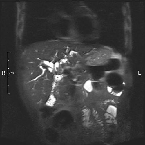
A 62-year-old man who had undergone Roux-en-Y hepaticojejunostomy because of choledochal cyst presented with jaundice. Magnetic resonance cholangiopancreatography revealed obstructive stones in left intrahepatic duct (Fig. 1).
Double-ballon enteroscope-assisted endoscopic retrograde cholangiopancreatography was initially attempted but failed to reach anastomosis. Endoscopic ultrasound-guided hepaticogastrostomy (HGS) was alternatively performed (Fig. 2a). Cholangiography confirmed multiple intrahepatic bile duct stones and a 2 cm length anastomotic stricture. After dilatation of the stomach wall and anastomotic stricture, a 10 × 80 mm fully covered self-expanding metal stent (FCSEMS) was deployed.
One week later when the FCSEMS was assumed to be fully expanded (Fig. 2b), transluminal stone removal was attempted. With the concern of stent migration during stone removal, a clip and dental floss was used for FCSEMS traction and fixation (Fig. 2c). SpyGlass-guided laser lithotripsy and stone extraction was performed through the endoscopic ultrasound-guided HGS route (Fig. 2d). A coaxial double-pigtail plastic stent was inserted through the FCSEMS to function as an anchor. No postoperative adverse event was observed. Jaundice was rapidly relieved (Video S1).
Removal of left intrahepatic duct stones via the HGS route has been described in sporadic cases.1 Three highlights of this case are specifically introduced as follows. First, given that the stone's size exceeded the width of intrahepatic bile duct and the diameter of metal stent when it was not fully expanded, adequate stone removal cannot be accomplished in a single session. An HGS route must first be established. Lithotripsy was then carried out. Second, to decrease the risk of stent migration during accessory devices pass-by, we borrowed the method of dental floss traction applied in endoscopic submucosal dissection.2 Last, anastomotic stenosis is difficult to alleviate from a single dilation. With the help of the HGS route, double-ballon enteroscope-assisted endoscopic retrograde cholangiopancreatography can be easily performed by rendezvous technique in the subsequent session and the transgastric stent can be removed thereby.
Authors declare no conflict of interest for this article.
This study was supported by the National Natural Science Foundation of China (Grant No. 82070663 [J.Y.M.]) and Specific Research Fund of The Innovation Platform for Academicians of Hainan Province (Grant No. YSPTZX202029 [K.X.W.]).
期刊介绍:
Digestive Endoscopy (DEN) is the official journal of the Japan Gastroenterological Endoscopy Society, the Asian Pacific Society for Digestive Endoscopy and the World Endoscopy Organization. Digestive Endoscopy serves as a medium for presenting original articles that offer significant contributions to knowledge in the broad field of endoscopy. The Journal also includes Reviews, Original Articles, How I Do It, Case Reports (only of exceptional interest and novelty are accepted), Letters, Techniques and Images, abstracts and news items that may be of interest to endoscopists.

 求助内容:
求助内容: 应助结果提醒方式:


