Patrícia Neto-Fernandes, Clara Chamadoira, Carolina Silva, Leila Pereira, Rui Vaz, Manuel Rito, Manuel J Ferreira-Pinto
{"title":"术中三维透视可准确预测脑深部刺激手术的最终电极位置。","authors":"Patrícia Neto-Fernandes, Clara Chamadoira, Carolina Silva, Leila Pereira, Rui Vaz, Manuel Rito, Manuel J Ferreira-Pinto","doi":"10.1007/s00701-024-06214-8","DOIUrl":null,"url":null,"abstract":"<p><strong>Purpose: </strong>In the absence of an intraoperative CT or MRI setup, post-implantation confirmation of electrode position in deep brain stimulation (DBS) requires patient transportation to the radiology unit, prolonging surgery time. This project aims to validate intraoperative 3D fluoroscopy (3DF), a widely available tool in Neurosurgical units, as a method to determine final electrode position.</p><p><strong>Methods: </strong>We performed a retrospective study including 64 patients (124 electrodes) who underwent DBS at our institution. Intraoperative 3DF after electrode implantation and postoperative volumetric CT were acquired. The Euclidean coordinates of the electrode tip displayed in both imaging modalities were determined and inter-method deviations were assessed. Pneumocephalus was quantified and its potential impact in determining the electrode position analyzed. Finally, 3DF and CT-imposed exposure to radiation was compared.</p><p><strong>Results: </strong>The difference between the electrode tip estimated by 3DF and CT was 0.85 ± 0.03 mm, and not significantly different (p = 0.11 for the distance to MCP assessed by both methods), but was, instead, highly correlated (p = 0.91; p < 0.0001). Even though pneumocephalus was larger in 3DF (6.89 ± 1.76 vs 5.18 ± 1.37 mm<sup>3</sup> in the CT group, p < 0.001), it was not correlated with the difference in electrode position measured by both techniques (p = 0.17; p = 0.06). Radiation exposure from 3DF is significantly lower than CT (0.36 ± 0.03 vs 2.08 ± 0.05 mSv; p < 0.0001).</p><p><strong>Conclusions: </strong>Intraoperative 3DF is comparable to CT in determining the final DBS electrode position. Being a method with fewer radiation exposure, less expensive, faster and that avoids patient transportation outside the operation room, it is a valid tool to replace postoperative CT.</p>","PeriodicalId":7370,"journal":{"name":"Acta Neurochirurgica","volume":null,"pages":null},"PeriodicalIF":1.9000,"publicationDate":"2024-08-07","publicationTypes":"Journal Article","fieldsOfStudy":null,"isOpenAccess":false,"openAccessPdf":"https://www.ncbi.nlm.nih.gov/pmc/articles/PMC11303432/pdf/","citationCount":"0","resultStr":"{\"title\":\"Intraoperative 3D fluoroscopy accurately predicts final electrode position in deep brain stimulation surgery.\",\"authors\":\"Patrícia Neto-Fernandes, Clara Chamadoira, Carolina Silva, Leila Pereira, Rui Vaz, Manuel Rito, Manuel J Ferreira-Pinto\",\"doi\":\"10.1007/s00701-024-06214-8\",\"DOIUrl\":null,\"url\":null,\"abstract\":\"<p><strong>Purpose: </strong>In the absence of an intraoperative CT or MRI setup, post-implantation confirmation of electrode position in deep brain stimulation (DBS) requires patient transportation to the radiology unit, prolonging surgery time. This project aims to validate intraoperative 3D fluoroscopy (3DF), a widely available tool in Neurosurgical units, as a method to determine final electrode position.</p><p><strong>Methods: </strong>We performed a retrospective study including 64 patients (124 electrodes) who underwent DBS at our institution. Intraoperative 3DF after electrode implantation and postoperative volumetric CT were acquired. The Euclidean coordinates of the electrode tip displayed in both imaging modalities were determined and inter-method deviations were assessed. Pneumocephalus was quantified and its potential impact in determining the electrode position analyzed. Finally, 3DF and CT-imposed exposure to radiation was compared.</p><p><strong>Results: </strong>The difference between the electrode tip estimated by 3DF and CT was 0.85 ± 0.03 mm, and not significantly different (p = 0.11 for the distance to MCP assessed by both methods), but was, instead, highly correlated (p = 0.91; p < 0.0001). Even though pneumocephalus was larger in 3DF (6.89 ± 1.76 vs 5.18 ± 1.37 mm<sup>3</sup> in the CT group, p < 0.001), it was not correlated with the difference in electrode position measured by both techniques (p = 0.17; p = 0.06). Radiation exposure from 3DF is significantly lower than CT (0.36 ± 0.03 vs 2.08 ± 0.05 mSv; p < 0.0001).</p><p><strong>Conclusions: </strong>Intraoperative 3DF is comparable to CT in determining the final DBS electrode position. Being a method with fewer radiation exposure, less expensive, faster and that avoids patient transportation outside the operation room, it is a valid tool to replace postoperative CT.</p>\",\"PeriodicalId\":7370,\"journal\":{\"name\":\"Acta Neurochirurgica\",\"volume\":null,\"pages\":null},\"PeriodicalIF\":1.9000,\"publicationDate\":\"2024-08-07\",\"publicationTypes\":\"Journal Article\",\"fieldsOfStudy\":null,\"isOpenAccess\":false,\"openAccessPdf\":\"https://www.ncbi.nlm.nih.gov/pmc/articles/PMC11303432/pdf/\",\"citationCount\":\"0\",\"resultStr\":null,\"platform\":\"Semanticscholar\",\"paperid\":null,\"PeriodicalName\":\"Acta Neurochirurgica\",\"FirstCategoryId\":\"3\",\"ListUrlMain\":\"https://doi.org/10.1007/s00701-024-06214-8\",\"RegionNum\":3,\"RegionCategory\":\"医学\",\"ArticlePicture\":[],\"TitleCN\":null,\"AbstractTextCN\":null,\"PMCID\":null,\"EPubDate\":\"\",\"PubModel\":\"\",\"JCR\":\"Q3\",\"JCRName\":\"CLINICAL NEUROLOGY\",\"Score\":null,\"Total\":0}","platform":"Semanticscholar","paperid":null,"PeriodicalName":"Acta Neurochirurgica","FirstCategoryId":"3","ListUrlMain":"https://doi.org/10.1007/s00701-024-06214-8","RegionNum":3,"RegionCategory":"医学","ArticlePicture":[],"TitleCN":null,"AbstractTextCN":null,"PMCID":null,"EPubDate":"","PubModel":"","JCR":"Q3","JCRName":"CLINICAL NEUROLOGY","Score":null,"Total":0}
Intraoperative 3D fluoroscopy accurately predicts final electrode position in deep brain stimulation surgery.
Purpose: In the absence of an intraoperative CT or MRI setup, post-implantation confirmation of electrode position in deep brain stimulation (DBS) requires patient transportation to the radiology unit, prolonging surgery time. This project aims to validate intraoperative 3D fluoroscopy (3DF), a widely available tool in Neurosurgical units, as a method to determine final electrode position.
Methods: We performed a retrospective study including 64 patients (124 electrodes) who underwent DBS at our institution. Intraoperative 3DF after electrode implantation and postoperative volumetric CT were acquired. The Euclidean coordinates of the electrode tip displayed in both imaging modalities were determined and inter-method deviations were assessed. Pneumocephalus was quantified and its potential impact in determining the electrode position analyzed. Finally, 3DF and CT-imposed exposure to radiation was compared.
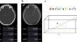
Results: The difference between the electrode tip estimated by 3DF and CT was 0.85 ± 0.03 mm, and not significantly different (p = 0.11 for the distance to MCP assessed by both methods), but was, instead, highly correlated (p = 0.91; p < 0.0001). Even though pneumocephalus was larger in 3DF (6.89 ± 1.76 vs 5.18 ± 1.37 mm3 in the CT group, p < 0.001), it was not correlated with the difference in electrode position measured by both techniques (p = 0.17; p = 0.06). Radiation exposure from 3DF is significantly lower than CT (0.36 ± 0.03 vs 2.08 ± 0.05 mSv; p < 0.0001).
Conclusions: Intraoperative 3DF is comparable to CT in determining the final DBS electrode position. Being a method with fewer radiation exposure, less expensive, faster and that avoids patient transportation outside the operation room, it is a valid tool to replace postoperative CT.
期刊介绍:
The journal "Acta Neurochirurgica" publishes only original papers useful both to research and clinical work. Papers should deal with clinical neurosurgery - diagnosis and diagnostic techniques, operative surgery and results, postoperative treatment - or with research work in neuroscience if the underlying questions or the results are of neurosurgical interest. Reports on congresses are given in brief accounts. As official organ of the European Association of Neurosurgical Societies the journal publishes all announcements of the E.A.N.S. and reports on the activities of its member societies. Only contributions written in English will be accepted.

 求助内容:
求助内容: 应助结果提醒方式:


