{"title":"利用磁共振成像和几何特征评估 MCI 和 AD 的穹窿结构变化","authors":"Ahsan Ali, Jac Fredo Agastinose Ronickom, Ramakrishnan Swaminathan","doi":"10.1007/s40846-024-00883-7","DOIUrl":null,"url":null,"abstract":"<h3 data-test=\"abstract-sub-heading\">Purpose</h3><p>Mild cognitive impairment (MCI) and Alzheimer’s disease (AD) are known to cause geometrical changes in the integrity of the fornix, which plays a crucial role in memory formation and retrieval. The objective of this study is to analyse structural variations in the fornix region using structural magnetic resonance (sMR) images and geometrical features.</p><h3 data-test=\"abstract-sub-heading\">Methods</h3><p>Initially, the fornix region of the brain is segmented from the sMR images of normal cognitive (NC), MCI and AD using the FreeSurfer software package. Further, geometrical features such as volume, equivalent diameter, extent, major axis length, and solidity are extracted to investigate the changes in the structure of the fornix in MCI and AD conditions.</p><h3 data-test=\"abstract-sub-heading\">Results</h3><p>The segmentation results show that FreeSurfer software is able to delineate the irregular boundaries of the fornix region accurately. The extent, major axis length, and solidity features are found to be statistically significant (<i>p</i> < 0.001) in discriminating NC, MCI and AD. It indicates that the considered features can capture the geometrical variation in the fornix structure.</p><h3 data-test=\"abstract-sub-heading\">Conclusion</h3><p>The reported approach can facilitate the early diagnosis of the disease, as the distinction of AD in the preclinical stage is complex and clinically significant.</p>","PeriodicalId":50133,"journal":{"name":"Journal of Medical and Biological Engineering","volume":"45 1","pages":""},"PeriodicalIF":1.6000,"publicationDate":"2024-07-29","publicationTypes":"Journal Article","fieldsOfStudy":null,"isOpenAccess":false,"openAccessPdf":"","citationCount":"0","resultStr":"{\"title\":\"Assessment of Structural Variations in Fornix of MCI and AD Using MR Images and Geometrical Features\",\"authors\":\"Ahsan Ali, Jac Fredo Agastinose Ronickom, Ramakrishnan Swaminathan\",\"doi\":\"10.1007/s40846-024-00883-7\",\"DOIUrl\":null,\"url\":null,\"abstract\":\"<h3 data-test=\\\"abstract-sub-heading\\\">Purpose</h3><p>Mild cognitive impairment (MCI) and Alzheimer’s disease (AD) are known to cause geometrical changes in the integrity of the fornix, which plays a crucial role in memory formation and retrieval. The objective of this study is to analyse structural variations in the fornix region using structural magnetic resonance (sMR) images and geometrical features.</p><h3 data-test=\\\"abstract-sub-heading\\\">Methods</h3><p>Initially, the fornix region of the brain is segmented from the sMR images of normal cognitive (NC), MCI and AD using the FreeSurfer software package. Further, geometrical features such as volume, equivalent diameter, extent, major axis length, and solidity are extracted to investigate the changes in the structure of the fornix in MCI and AD conditions.</p><h3 data-test=\\\"abstract-sub-heading\\\">Results</h3><p>The segmentation results show that FreeSurfer software is able to delineate the irregular boundaries of the fornix region accurately. The extent, major axis length, and solidity features are found to be statistically significant (<i>p</i> < 0.001) in discriminating NC, MCI and AD. It indicates that the considered features can capture the geometrical variation in the fornix structure.</p><h3 data-test=\\\"abstract-sub-heading\\\">Conclusion</h3><p>The reported approach can facilitate the early diagnosis of the disease, as the distinction of AD in the preclinical stage is complex and clinically significant.</p>\",\"PeriodicalId\":50133,\"journal\":{\"name\":\"Journal of Medical and Biological Engineering\",\"volume\":\"45 1\",\"pages\":\"\"},\"PeriodicalIF\":1.6000,\"publicationDate\":\"2024-07-29\",\"publicationTypes\":\"Journal Article\",\"fieldsOfStudy\":null,\"isOpenAccess\":false,\"openAccessPdf\":\"\",\"citationCount\":\"0\",\"resultStr\":null,\"platform\":\"Semanticscholar\",\"paperid\":null,\"PeriodicalName\":\"Journal of Medical and Biological Engineering\",\"FirstCategoryId\":\"5\",\"ListUrlMain\":\"https://doi.org/10.1007/s40846-024-00883-7\",\"RegionNum\":4,\"RegionCategory\":\"医学\",\"ArticlePicture\":[],\"TitleCN\":null,\"AbstractTextCN\":null,\"PMCID\":null,\"EPubDate\":\"\",\"PubModel\":\"\",\"JCR\":\"Q4\",\"JCRName\":\"ENGINEERING, BIOMEDICAL\",\"Score\":null,\"Total\":0}","platform":"Semanticscholar","paperid":null,"PeriodicalName":"Journal of Medical and Biological Engineering","FirstCategoryId":"5","ListUrlMain":"https://doi.org/10.1007/s40846-024-00883-7","RegionNum":4,"RegionCategory":"医学","ArticlePicture":[],"TitleCN":null,"AbstractTextCN":null,"PMCID":null,"EPubDate":"","PubModel":"","JCR":"Q4","JCRName":"ENGINEERING, BIOMEDICAL","Score":null,"Total":0}
引用次数: 0
摘要
目的众所周知,轻度认知障碍(MCI)和阿尔茨海默病(AD)会导致穹窿的完整性发生几何变化,而穹窿在记忆的形成和检索中起着至关重要的作用。本研究的目的是利用结构磁共振(sMR)图像和几何特征来分析穹窿区的结构变化。方法首先,使用 FreeSurfer 软件包从正常认知(NC)、MCI 和 AD 的 sMR 图像中分割出大脑的穹窿区。结果分割结果表明,FreeSurfer 软件能准确划分出穹窿区域的不规则边界。在区分 NC、MCI 和 AD 时,发现范围、主轴长度和稳固性特征具有统计学意义(p < 0.001)。结论:由于临床前阶段的 AD 区分复杂且具有临床意义,因此所报告的方法有助于疾病的早期诊断。
Assessment of Structural Variations in Fornix of MCI and AD Using MR Images and Geometrical Features
Purpose
Mild cognitive impairment (MCI) and Alzheimer’s disease (AD) are known to cause geometrical changes in the integrity of the fornix, which plays a crucial role in memory formation and retrieval. The objective of this study is to analyse structural variations in the fornix region using structural magnetic resonance (sMR) images and geometrical features.
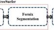
Methods
Initially, the fornix region of the brain is segmented from the sMR images of normal cognitive (NC), MCI and AD using the FreeSurfer software package. Further, geometrical features such as volume, equivalent diameter, extent, major axis length, and solidity are extracted to investigate the changes in the structure of the fornix in MCI and AD conditions.
Results
The segmentation results show that FreeSurfer software is able to delineate the irregular boundaries of the fornix region accurately. The extent, major axis length, and solidity features are found to be statistically significant (p < 0.001) in discriminating NC, MCI and AD. It indicates that the considered features can capture the geometrical variation in the fornix structure.
Conclusion
The reported approach can facilitate the early diagnosis of the disease, as the distinction of AD in the preclinical stage is complex and clinically significant.
期刊介绍:
The purpose of Journal of Medical and Biological Engineering, JMBE, is committed to encouraging and providing the standard of biomedical engineering. The journal is devoted to publishing papers related to clinical engineering, biomedical signals, medical imaging, bio-informatics, tissue engineering, and so on. Other than the above articles, any contributions regarding hot issues and technological developments that help reach the purpose are also included.

 求助内容:
求助内容: 应助结果提醒方式:


