Yasmin Bay , Federico Javier Miguez Cabello , Chloe C. Koens , Stine M. Frantsen , Darryl S. Pickering , Karla Frydenvang , Pierre Francotte , Bernard Pirotte , Anders S. Kristensen , Derek Bowie , Jette Sandholm Kastrup
{"title":"GluK1 配体结合结构域与凯恩酸盐和全跨度正异构调节剂 BPAM538 的晶体结构。","authors":"Yasmin Bay , Federico Javier Miguez Cabello , Chloe C. Koens , Stine M. Frantsen , Darryl S. Pickering , Karla Frydenvang , Pierre Francotte , Bernard Pirotte , Anders S. Kristensen , Derek Bowie , Jette Sandholm Kastrup","doi":"10.1016/j.jsb.2024.108113","DOIUrl":null,"url":null,"abstract":"<div><p>Kainate receptors play an important role in the central nervous system by mediating postsynaptic excitatory neurotransmission and modulating the release of the inhibitory neurotransmitter GABA through a presynaptic mechanism. To date, only three structures of the ligand-binding domain (LBD) of the kainate receptor subunit GluK1 in complex with positive allosteric modulators have been determined by X-ray crystallography, all belonging to class II modulators. Here, we report a high-resolution structure of GluK1-LBD in complex with kainate and BPAM538, which belongs to the full-spanning class III. One BPAM538 molecule binds at the GluK1 dimer interface, thereby occupying two allosteric binding sites simultaneously. BPAM538 stabilizes the active receptor conformation with only minor conformational changes being introduced to the receptor. Using a calcium-sensitive fluorescence-based assay, a 5-fold potentiation of the kainate response (100 μM) was observed in presence of 100 μM BPAM538 at GluK1(<em>Q</em>)<sub>b</sub>, whereas no potentiation was observed at GluK2(<em>VCQ</em>)<sub>a</sub>. Using electrophysiology recordings of outside-out patches excised from HEK293 cells, BPAM538 increased the peak response of GluK1(<em>Q</em>)<sub>b</sub> co-expressed with NETO2 to rapid application of 10 mM L-glutamate with 130 ± 20 %, and decreased desensitization determined as the steady-state/peak response ratio from 23 ± 2 % to 90 ± 4 %. Based on dose–response relationship experiments on GluK1(<em>Q</em>)<sub>b</sub> the EC<sub>50</sub> of BPAM538 was estimated to be 58 ± 29 μM.</p></div>","PeriodicalId":17074,"journal":{"name":"Journal of structural biology","volume":"216 3","pages":"Article 108113"},"PeriodicalIF":2.7000,"publicationDate":"2024-07-28","publicationTypes":"Journal Article","fieldsOfStudy":null,"isOpenAccess":false,"openAccessPdf":"https://www.sciencedirect.com/science/article/pii/S1047847724000534/pdfft?md5=fd36f2c650fe072db6eace68cdb0888f&pid=1-s2.0-S1047847724000534-main.pdf","citationCount":"0","resultStr":"{\"title\":\"Crystal structure of the GluK1 ligand-binding domain with kainate and the full-spanning positive allosteric modulator BPAM538\",\"authors\":\"Yasmin Bay , Federico Javier Miguez Cabello , Chloe C. Koens , Stine M. Frantsen , Darryl S. Pickering , Karla Frydenvang , Pierre Francotte , Bernard Pirotte , Anders S. Kristensen , Derek Bowie , Jette Sandholm Kastrup\",\"doi\":\"10.1016/j.jsb.2024.108113\",\"DOIUrl\":null,\"url\":null,\"abstract\":\"<div><p>Kainate receptors play an important role in the central nervous system by mediating postsynaptic excitatory neurotransmission and modulating the release of the inhibitory neurotransmitter GABA through a presynaptic mechanism. To date, only three structures of the ligand-binding domain (LBD) of the kainate receptor subunit GluK1 in complex with positive allosteric modulators have been determined by X-ray crystallography, all belonging to class II modulators. Here, we report a high-resolution structure of GluK1-LBD in complex with kainate and BPAM538, which belongs to the full-spanning class III. One BPAM538 molecule binds at the GluK1 dimer interface, thereby occupying two allosteric binding sites simultaneously. BPAM538 stabilizes the active receptor conformation with only minor conformational changes being introduced to the receptor. Using a calcium-sensitive fluorescence-based assay, a 5-fold potentiation of the kainate response (100 μM) was observed in presence of 100 μM BPAM538 at GluK1(<em>Q</em>)<sub>b</sub>, whereas no potentiation was observed at GluK2(<em>VCQ</em>)<sub>a</sub>. Using electrophysiology recordings of outside-out patches excised from HEK293 cells, BPAM538 increased the peak response of GluK1(<em>Q</em>)<sub>b</sub> co-expressed with NETO2 to rapid application of 10 mM L-glutamate with 130 ± 20 %, and decreased desensitization determined as the steady-state/peak response ratio from 23 ± 2 % to 90 ± 4 %. Based on dose–response relationship experiments on GluK1(<em>Q</em>)<sub>b</sub> the EC<sub>50</sub> of BPAM538 was estimated to be 58 ± 29 μM.</p></div>\",\"PeriodicalId\":17074,\"journal\":{\"name\":\"Journal of structural biology\",\"volume\":\"216 3\",\"pages\":\"Article 108113\"},\"PeriodicalIF\":2.7000,\"publicationDate\":\"2024-07-28\",\"publicationTypes\":\"Journal Article\",\"fieldsOfStudy\":null,\"isOpenAccess\":false,\"openAccessPdf\":\"https://www.sciencedirect.com/science/article/pii/S1047847724000534/pdfft?md5=fd36f2c650fe072db6eace68cdb0888f&pid=1-s2.0-S1047847724000534-main.pdf\",\"citationCount\":\"0\",\"resultStr\":null,\"platform\":\"Semanticscholar\",\"paperid\":null,\"PeriodicalName\":\"Journal of structural biology\",\"FirstCategoryId\":\"99\",\"ListUrlMain\":\"https://www.sciencedirect.com/science/article/pii/S1047847724000534\",\"RegionNum\":3,\"RegionCategory\":\"生物学\",\"ArticlePicture\":[],\"TitleCN\":null,\"AbstractTextCN\":null,\"PMCID\":null,\"EPubDate\":\"\",\"PubModel\":\"\",\"JCR\":\"Q3\",\"JCRName\":\"BIOCHEMISTRY & MOLECULAR BIOLOGY\",\"Score\":null,\"Total\":0}","platform":"Semanticscholar","paperid":null,"PeriodicalName":"Journal of structural biology","FirstCategoryId":"99","ListUrlMain":"https://www.sciencedirect.com/science/article/pii/S1047847724000534","RegionNum":3,"RegionCategory":"生物学","ArticlePicture":[],"TitleCN":null,"AbstractTextCN":null,"PMCID":null,"EPubDate":"","PubModel":"","JCR":"Q3","JCRName":"BIOCHEMISTRY & MOLECULAR BIOLOGY","Score":null,"Total":0}
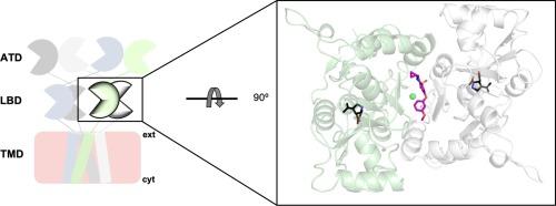
Crystal structure of the GluK1 ligand-binding domain with kainate and the full-spanning positive allosteric modulator BPAM538
Kainate receptors play an important role in the central nervous system by mediating postsynaptic excitatory neurotransmission and modulating the release of the inhibitory neurotransmitter GABA through a presynaptic mechanism. To date, only three structures of the ligand-binding domain (LBD) of the kainate receptor subunit GluK1 in complex with positive allosteric modulators have been determined by X-ray crystallography, all belonging to class II modulators. Here, we report a high-resolution structure of GluK1-LBD in complex with kainate and BPAM538, which belongs to the full-spanning class III. One BPAM538 molecule binds at the GluK1 dimer interface, thereby occupying two allosteric binding sites simultaneously. BPAM538 stabilizes the active receptor conformation with only minor conformational changes being introduced to the receptor. Using a calcium-sensitive fluorescence-based assay, a 5-fold potentiation of the kainate response (100 μM) was observed in presence of 100 μM BPAM538 at GluK1(Q)b, whereas no potentiation was observed at GluK2(VCQ)a. Using electrophysiology recordings of outside-out patches excised from HEK293 cells, BPAM538 increased the peak response of GluK1(Q)b co-expressed with NETO2 to rapid application of 10 mM L-glutamate with 130 ± 20 %, and decreased desensitization determined as the steady-state/peak response ratio from 23 ± 2 % to 90 ± 4 %. Based on dose–response relationship experiments on GluK1(Q)b the EC50 of BPAM538 was estimated to be 58 ± 29 μM.
期刊介绍:
Journal of Structural Biology (JSB) has an open access mirror journal, the Journal of Structural Biology: X (JSBX), sharing the same aims and scope, editorial team, submission system and rigorous peer review. Since both journals share the same editorial system, you may submit your manuscript via either journal homepage. You will be prompted during submission (and revision) to choose in which to publish your article. The editors and reviewers are not aware of the choice you made until the article has been published online. JSB and JSBX publish papers dealing with the structural analysis of living material at every level of organization by all methods that lead to an understanding of biological function in terms of molecular and supermolecular structure.
Techniques covered include:
• Light microscopy including confocal microscopy
• All types of electron microscopy
• X-ray diffraction
• Nuclear magnetic resonance
• Scanning force microscopy, scanning probe microscopy, and tunneling microscopy
• Digital image processing
• Computational insights into structure

 求助内容:
求助内容: 应助结果提醒方式:


