Kai Zhao, Yapeng Wang, Dong Li, Yanping Ding, Ying Yang, Qudong Yin, Yunhong Ma
{"title":"植入 S1AIS 作为骶髂关节固定技术具有优先权。","authors":"Kai Zhao, Yapeng Wang, Dong Li, Yanping Ding, Ying Yang, Qudong Yin, Yunhong Ma","doi":"10.1007/s00586-024-08423-2","DOIUrl":null,"url":null,"abstract":"<p><strong>Purpose: </strong>The sacral alar-iliac screw (SAIS) fixation technique has evolved from spinopelvic fixation which originated from S2AIS to sacroiliac joint fixation, with more reports regarding its application of S2AIS than S1AIS. However, there is a lack of comparative evidence to determine which technique is superior for sacroiliac joint fixation. This study aimed to determine which of the screws was superior in terms of implantation safety and biomechanical stability for sacroiliac joint fixation.</p><p><strong>Methods: </strong>CT data of 80 normal pelvises were analyzed to measure the insertable range, trajectory lengths and widths of both S1AIS and S2AIS on 3D reconstruction models. Φ 6.5 mm and 8.0 mm screws were implanted on the left and right sides of fifty 3D printed pelvic models respectively to observe for breach of screw implantation. Ten synthetic pelvis models were used to simulate type C Tile injuries, and divided into 2 groups with an anterior plate and posterior fixation using one S1AIS or S2AIS on each side. The stiffness and maximum load of the plated and fixated models were measured under vertical loading.</p><p><strong>Results: </strong>The trajectory lengths and widths of the S1AIS and S2AIS were similar (p > 0.05) and there was no breach for Φ 6.5 mm SAIS. However, both the insertable range and trajectory length on the sacral side of S2AIS (234.56 ± 10.06 mm<sup>2</sup>, 40.97 ± 2.81 mm) were significantly less, and the breach rate of the posterior lateral cortex of the Φ 8.0 mm S2AIS (46%) was significantly higher than the S1AIS (307.55 ± 10.42 mm<sup>2</sup>, 42.16 ± 3.06 mm, and 2%, p < 0.05). The stiffness and maximum load of S2AIS were less than S1AIS but the difference was not statistically significant (p > 0.05).</p><p><strong>Conclusion: </strong>S1AIS and S2AIS have similar screw trajectories and stability. However, S1AIS has a larger insertable range, less breach of the posterior lateral sacral cortex and longer trajectory length on the sacral side than S2AIS, which indicates S1AIS has higher implantation safety and a trend of better mechanical performance over S2AIS for sacroiliac joint fixation. Furthermore, S2AIS with an excessively large diameter should be used with caution for sacroiliac joint fixation.</p>","PeriodicalId":12323,"journal":{"name":"European Spine Journal","volume":null,"pages":null},"PeriodicalIF":2.6000,"publicationDate":"2024-09-01","publicationTypes":"Journal Article","fieldsOfStudy":null,"isOpenAccess":false,"openAccessPdf":"","citationCount":"0","resultStr":"{\"title\":\"Implantation of S1AIS has priority as a sacroiliac joint fixation technique.\",\"authors\":\"Kai Zhao, Yapeng Wang, Dong Li, Yanping Ding, Ying Yang, Qudong Yin, Yunhong Ma\",\"doi\":\"10.1007/s00586-024-08423-2\",\"DOIUrl\":null,\"url\":null,\"abstract\":\"<p><strong>Purpose: </strong>The sacral alar-iliac screw (SAIS) fixation technique has evolved from spinopelvic fixation which originated from S2AIS to sacroiliac joint fixation, with more reports regarding its application of S2AIS than S1AIS. However, there is a lack of comparative evidence to determine which technique is superior for sacroiliac joint fixation. This study aimed to determine which of the screws was superior in terms of implantation safety and biomechanical stability for sacroiliac joint fixation.</p><p><strong>Methods: </strong>CT data of 80 normal pelvises were analyzed to measure the insertable range, trajectory lengths and widths of both S1AIS and S2AIS on 3D reconstruction models. Φ 6.5 mm and 8.0 mm screws were implanted on the left and right sides of fifty 3D printed pelvic models respectively to observe for breach of screw implantation. Ten synthetic pelvis models were used to simulate type C Tile injuries, and divided into 2 groups with an anterior plate and posterior fixation using one S1AIS or S2AIS on each side. The stiffness and maximum load of the plated and fixated models were measured under vertical loading.</p><p><strong>Results: </strong>The trajectory lengths and widths of the S1AIS and S2AIS were similar (p > 0.05) and there was no breach for Φ 6.5 mm SAIS. However, both the insertable range and trajectory length on the sacral side of S2AIS (234.56 ± 10.06 mm<sup>2</sup>, 40.97 ± 2.81 mm) were significantly less, and the breach rate of the posterior lateral cortex of the Φ 8.0 mm S2AIS (46%) was significantly higher than the S1AIS (307.55 ± 10.42 mm<sup>2</sup>, 42.16 ± 3.06 mm, and 2%, p < 0.05). The stiffness and maximum load of S2AIS were less than S1AIS but the difference was not statistically significant (p > 0.05).</p><p><strong>Conclusion: </strong>S1AIS and S2AIS have similar screw trajectories and stability. However, S1AIS has a larger insertable range, less breach of the posterior lateral sacral cortex and longer trajectory length on the sacral side than S2AIS, which indicates S1AIS has higher implantation safety and a trend of better mechanical performance over S2AIS for sacroiliac joint fixation. Furthermore, S2AIS with an excessively large diameter should be used with caution for sacroiliac joint fixation.</p>\",\"PeriodicalId\":12323,\"journal\":{\"name\":\"European Spine Journal\",\"volume\":null,\"pages\":null},\"PeriodicalIF\":2.6000,\"publicationDate\":\"2024-09-01\",\"publicationTypes\":\"Journal Article\",\"fieldsOfStudy\":null,\"isOpenAccess\":false,\"openAccessPdf\":\"\",\"citationCount\":\"0\",\"resultStr\":null,\"platform\":\"Semanticscholar\",\"paperid\":null,\"PeriodicalName\":\"European Spine Journal\",\"FirstCategoryId\":\"3\",\"ListUrlMain\":\"https://doi.org/10.1007/s00586-024-08423-2\",\"RegionNum\":3,\"RegionCategory\":\"医学\",\"ArticlePicture\":[],\"TitleCN\":null,\"AbstractTextCN\":null,\"PMCID\":null,\"EPubDate\":\"2024/7/29 0:00:00\",\"PubModel\":\"Epub\",\"JCR\":\"Q2\",\"JCRName\":\"CLINICAL NEUROLOGY\",\"Score\":null,\"Total\":0}","platform":"Semanticscholar","paperid":null,"PeriodicalName":"European Spine Journal","FirstCategoryId":"3","ListUrlMain":"https://doi.org/10.1007/s00586-024-08423-2","RegionNum":3,"RegionCategory":"医学","ArticlePicture":[],"TitleCN":null,"AbstractTextCN":null,"PMCID":null,"EPubDate":"2024/7/29 0:00:00","PubModel":"Epub","JCR":"Q2","JCRName":"CLINICAL NEUROLOGY","Score":null,"Total":0}
Implantation of S1AIS has priority as a sacroiliac joint fixation technique.
Purpose: The sacral alar-iliac screw (SAIS) fixation technique has evolved from spinopelvic fixation which originated from S2AIS to sacroiliac joint fixation, with more reports regarding its application of S2AIS than S1AIS. However, there is a lack of comparative evidence to determine which technique is superior for sacroiliac joint fixation. This study aimed to determine which of the screws was superior in terms of implantation safety and biomechanical stability for sacroiliac joint fixation.
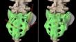
Methods: CT data of 80 normal pelvises were analyzed to measure the insertable range, trajectory lengths and widths of both S1AIS and S2AIS on 3D reconstruction models. Φ 6.5 mm and 8.0 mm screws were implanted on the left and right sides of fifty 3D printed pelvic models respectively to observe for breach of screw implantation. Ten synthetic pelvis models were used to simulate type C Tile injuries, and divided into 2 groups with an anterior plate and posterior fixation using one S1AIS or S2AIS on each side. The stiffness and maximum load of the plated and fixated models were measured under vertical loading.
Results: The trajectory lengths and widths of the S1AIS and S2AIS were similar (p > 0.05) and there was no breach for Φ 6.5 mm SAIS. However, both the insertable range and trajectory length on the sacral side of S2AIS (234.56 ± 10.06 mm2, 40.97 ± 2.81 mm) were significantly less, and the breach rate of the posterior lateral cortex of the Φ 8.0 mm S2AIS (46%) was significantly higher than the S1AIS (307.55 ± 10.42 mm2, 42.16 ± 3.06 mm, and 2%, p < 0.05). The stiffness and maximum load of S2AIS were less than S1AIS but the difference was not statistically significant (p > 0.05).
Conclusion: S1AIS and S2AIS have similar screw trajectories and stability. However, S1AIS has a larger insertable range, less breach of the posterior lateral sacral cortex and longer trajectory length on the sacral side than S2AIS, which indicates S1AIS has higher implantation safety and a trend of better mechanical performance over S2AIS for sacroiliac joint fixation. Furthermore, S2AIS with an excessively large diameter should be used with caution for sacroiliac joint fixation.
期刊介绍:
"European Spine Journal" is a publication founded in response to the increasing trend toward specialization in spinal surgery and spinal pathology in general. The Journal is devoted to all spine related disciplines, including functional and surgical anatomy of the spine, biomechanics and pathophysiology, diagnostic procedures, and neurology, surgery and outcomes. The aim of "European Spine Journal" is to support the further development of highly innovative spine treatments including but not restricted to surgery and to provide an integrated and balanced view of diagnostic, research and treatment procedures as well as outcomes that will enhance effective collaboration among specialists worldwide. The “European Spine Journal” also participates in education by means of videos, interactive meetings and the endorsement of educative efforts.
Official publication of EUROSPINE, The Spine Society of Europe

 求助内容:
求助内容: 应助结果提醒方式:


