F. R. S. Andrade, E. L. da Silva, A. D. Marinho, A. C. X. Oliveira, D. Sánchez-Porras, F. Bermejo-Casares, R. C. Montenegro, V. Carriel, H. S. A. Monteiro, R. J. B. Jorge
{"title":"一种新的 L929 成纤维细胞微组织三维模型揭示了红腹锦鸡毒液及其抗毒血清的作用。","authors":"F. R. S. Andrade, E. L. da Silva, A. D. Marinho, A. C. X. Oliveira, D. Sánchez-Porras, F. Bermejo-Casares, R. C. Montenegro, V. Carriel, H. S. A. Monteiro, R. J. B. Jorge","doi":"10.1007/s00204-024-03824-0","DOIUrl":null,"url":null,"abstract":"<div><p>In Brazil, around 80% of snakebites are caused by snakes of the genus Bothrops. A three-dimensional culture model was standardized and used to perform treatments with <i>Bothrops erythromelas</i> venom (<i>Be</i>V) and its antivenom (AV). The MRC-5 and L929 cell lines were cultured at increasing cell densities. Morphometric parameters were evaluated through images obtained from an inverted microscope: solidity, circularity, and Feret diameter. L929 microtissues (MT) showed better morphometric data, and thus they were used for further analysis. MT viability was assessed using the acridine orange and ethidium bromide staining method, which showed viable cells in the MT on days 5, 7, and 10 of cultivation. Histochemical and histological analyses were performed, including hematoxylin/eosin staining, which showed a good structure of the spheroids. Alcian blue staining revealed the presence of acid proteoglycans. Immunohistochemical analysis with ki-67 showed different patterns of cell proliferation. The MT were also subjected to pharmacological tests using the <i>Be</i>V, in the presence or absence of its AV. The results showed that the venom was not cytotoxic, but it caused morphological changes. The MT showed cell detachment, losing their structure. The antivenom was able to partially prevent the venom activities.</p></div>","PeriodicalId":8329,"journal":{"name":"Archives of Toxicology","volume":"98 10","pages":"3503 - 3512"},"PeriodicalIF":4.8000,"publicationDate":"2024-07-15","publicationTypes":"Journal Article","fieldsOfStudy":null,"isOpenAccess":false,"openAccessPdf":"","citationCount":"0","resultStr":"{\"title\":\"A new 3D model of L929 fibroblasts microtissues uncovers the effects of Bothrops erythromelas venom and its antivenom\",\"authors\":\"F. R. S. Andrade, E. L. da Silva, A. D. Marinho, A. C. X. Oliveira, D. Sánchez-Porras, F. Bermejo-Casares, R. C. Montenegro, V. Carriel, H. S. A. Monteiro, R. J. B. Jorge\",\"doi\":\"10.1007/s00204-024-03824-0\",\"DOIUrl\":null,\"url\":null,\"abstract\":\"<div><p>In Brazil, around 80% of snakebites are caused by snakes of the genus Bothrops. A three-dimensional culture model was standardized and used to perform treatments with <i>Bothrops erythromelas</i> venom (<i>Be</i>V) and its antivenom (AV). The MRC-5 and L929 cell lines were cultured at increasing cell densities. Morphometric parameters were evaluated through images obtained from an inverted microscope: solidity, circularity, and Feret diameter. L929 microtissues (MT) showed better morphometric data, and thus they were used for further analysis. MT viability was assessed using the acridine orange and ethidium bromide staining method, which showed viable cells in the MT on days 5, 7, and 10 of cultivation. Histochemical and histological analyses were performed, including hematoxylin/eosin staining, which showed a good structure of the spheroids. Alcian blue staining revealed the presence of acid proteoglycans. Immunohistochemical analysis with ki-67 showed different patterns of cell proliferation. The MT were also subjected to pharmacological tests using the <i>Be</i>V, in the presence or absence of its AV. The results showed that the venom was not cytotoxic, but it caused morphological changes. The MT showed cell detachment, losing their structure. The antivenom was able to partially prevent the venom activities.</p></div>\",\"PeriodicalId\":8329,\"journal\":{\"name\":\"Archives of Toxicology\",\"volume\":\"98 10\",\"pages\":\"3503 - 3512\"},\"PeriodicalIF\":4.8000,\"publicationDate\":\"2024-07-15\",\"publicationTypes\":\"Journal Article\",\"fieldsOfStudy\":null,\"isOpenAccess\":false,\"openAccessPdf\":\"\",\"citationCount\":\"0\",\"resultStr\":null,\"platform\":\"Semanticscholar\",\"paperid\":null,\"PeriodicalName\":\"Archives of Toxicology\",\"FirstCategoryId\":\"3\",\"ListUrlMain\":\"https://link.springer.com/article/10.1007/s00204-024-03824-0\",\"RegionNum\":2,\"RegionCategory\":\"医学\",\"ArticlePicture\":[],\"TitleCN\":null,\"AbstractTextCN\":null,\"PMCID\":null,\"EPubDate\":\"\",\"PubModel\":\"\",\"JCR\":\"Q1\",\"JCRName\":\"TOXICOLOGY\",\"Score\":null,\"Total\":0}","platform":"Semanticscholar","paperid":null,"PeriodicalName":"Archives of Toxicology","FirstCategoryId":"3","ListUrlMain":"https://link.springer.com/article/10.1007/s00204-024-03824-0","RegionNum":2,"RegionCategory":"医学","ArticlePicture":[],"TitleCN":null,"AbstractTextCN":null,"PMCID":null,"EPubDate":"","PubModel":"","JCR":"Q1","JCRName":"TOXICOLOGY","Score":null,"Total":0}
A new 3D model of L929 fibroblasts microtissues uncovers the effects of Bothrops erythromelas venom and its antivenom
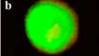
In Brazil, around 80% of snakebites are caused by snakes of the genus Bothrops. A three-dimensional culture model was standardized and used to perform treatments with Bothrops erythromelas venom (BeV) and its antivenom (AV). The MRC-5 and L929 cell lines were cultured at increasing cell densities. Morphometric parameters were evaluated through images obtained from an inverted microscope: solidity, circularity, and Feret diameter. L929 microtissues (MT) showed better morphometric data, and thus they were used for further analysis. MT viability was assessed using the acridine orange and ethidium bromide staining method, which showed viable cells in the MT on days 5, 7, and 10 of cultivation. Histochemical and histological analyses were performed, including hematoxylin/eosin staining, which showed a good structure of the spheroids. Alcian blue staining revealed the presence of acid proteoglycans. Immunohistochemical analysis with ki-67 showed different patterns of cell proliferation. The MT were also subjected to pharmacological tests using the BeV, in the presence or absence of its AV. The results showed that the venom was not cytotoxic, but it caused morphological changes. The MT showed cell detachment, losing their structure. The antivenom was able to partially prevent the venom activities.
期刊介绍:
Archives of Toxicology provides up-to-date information on the latest advances in toxicology. The journal places particular emphasis on studies relating to defined effects of chemicals and mechanisms of toxicity, including toxic activities at the molecular level, in humans and experimental animals. Coverage includes new insights into analysis and toxicokinetics and into forensic toxicology. Review articles of general interest to toxicologists are an additional important feature of the journal.
 求助内容:
求助内容: 应助结果提醒方式:


